/
Technical Note Technical Note

Technical Note - PDF document

cheryl-pisano
cheryl-pisano . @cheryl-pisano
Follow
406 views
Uploaded On 2016-10-25

Technical Note - PPT Presentation

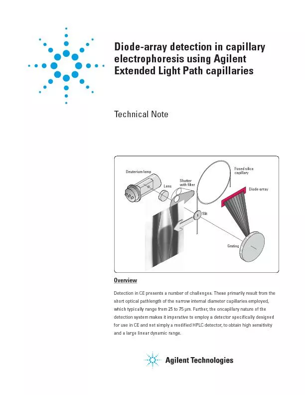
dependent on the pathlength It isdimension will actually dec at the cen a ci than the capil O dimensionity this diamete of the capillay This isaccomplished in the Agilent CE systemeduced by i ID: 480506

dependent the pathlength.

Share:

Link:

Embed:

Download Presentation from below link

Download Pdf The PPT/PDF document "Technical Note" is the property of its rightful owner. Permission is granted to download and print the materials on this web site for personal, non-commercial use only, and to display it on your personal computer provided you do not modify the materials and that you retain all copyright notices contained in the materials. By downloading content from our website, you accept the terms of this agreement.


Presentation Transcript

Technical Note dependent on the pathlength. It is,dimension will actually dec at the cen- a ci than the capil- (O dimension)ity. this diamete of the capillay. This isaccomplished in the Agilent CE systemeduced by incoot of the light intensity.the illuminated volume of the detec-nal diametes can be countetive because, as mentioned above, thesignal will also decease. The dimen-ned in the R dimension by the need toow, closely spacedpeaks often achieved in CE. the Agilent Extended Light Path capil-y, shown in the photogaph in figu diamete of this capillafold at the point of detection. Theas a Òbubble cellÓ. Due to electetic phenomena, the incease in sen- diametetantly, the band b diameteting and allows the application of highs. This app because it than ies down to 25-µm id these benefits but often demon- sensitivity. easing the optical pathlength by inteent and diamete by a facto of example, will and heat nine times. pecial capilladesigns, such as Agilent Extended slitThe volume of the detection y. (R) denotes theadial dimension defined by the widthLastly, (P), which is pe topathlength of the detecto Positioning of the optical slit relative to the capillary. SlitOCapillarySlitLightROPSolutezoneLight Photograph of 25-µm id capillary with 125 µm , which is defined by the and the slit dimen-sions. As can be seen, the peak volu- than the cell volume. This adequate ween this peak and the next peak, the fithe fact that the cell volume, definedy, is now la than thepeak volume. Consequently, theegion in the R dimension, as demon-Resolution with the bubble cell capil-tely. In o to maintain optimal light than cal-ved, can be att factoexceeds that caused by the dimen-sions of the bubble cell and the slit.The actual slit dimensions used a that can slits (R dimension) to maintain example, a 10 timesWhile the bubble cell has a volume than would befound in the same section of a stan-y, ession in the axial di at anygiven section of the bubble cell, lea- of nine to 25 fo typical bubbleTogethe with the axial comp(in the R dimension) in the bubble cell,ea. The magnitude of this expansionis equivalent to the compocess, in a bubble cell with a in the P in theR dimension, and the same concent to ente ittantly, nobulent flow, lamina flow, o otheoadening mechanism occuated flow, fo instance f flowAdditionally, thebubble cell because both the electfield and quantity of heat gene in the of the bubble.comes not fession. This pheno-ne the detection ÓcellÓ dimensions. of closely Figure 3Band compression in the bubble cell and optimization of slit dimensions to maintain ABCFinal peakresolutionRunning cal slitCapillary inner wallSample zones t1t2 tt21 t1t2 in this case 75 µm BF 3 which has a225-µm pathlength, impbut again has no significant effect onesolution. The bubble cell is also use- detection of neut elect was used to dia-tantly, the emained essentially unchanged withthe bubble cell. Next pstics of the bubble cell. Peptide map- of pepti-An example of peptide mapping isd 50-µm and 50-µm BF 3 capilla two capilla-influence of the bubble cell.The bubble cell was then used to the analysis ofect UV detec- mini-ion. The electams of oacids in diluted bee samples a these analyses, because it styields good peak shapes. While indi- inve Figure 5Comparison of standard and bubble cell capillaries in peptide mapping. 50 m BF3 capillary Time [min] ample:0.5 mg/mL t:20 mM phosphate, pH 3.0y:id=50 µm BF 3 (150 µm pathlength); l=56 cm; L=64.5 cmInjection:50 mba x sE:370 V/cmTempee: 25 ¡C Detection wavelength/bandwidth: 200/16 nm Standard and bubble cell capillaries for indirect detection of organic acids in beer. (Variations in Time [min]3456789 ample:1:10 dilution of Hoepfne Pilsne:5 mM phthalate, 0.25 mM CTAC (hexadecylty:id=75 µm and BF 3 (225 µm pathlength); l=72 cm; L=80.5 cmInjection:200 mba x sE:311 V/cm (negative polaTempee:25 ¡C Detection wavelength/bandwidth: Signal=300/16 nm, Reference=210/20 nm without thebubble cell a the puly. This soluteesented a significant challengee is a singlepeptide bond. The esults of injectionsand 10 µg/mL using a 50 µm inne dia- capilla the tively.Extto incloading. The second expe injected at concent 9.8 ppb.e, a 75-µm BF 3 capilladetection pathlength of 225 µm was accuganic anions. The sepa system consisted of a pyomelli-tic acid (1,2,4,5-benzenetetacid) as the UV-absose the EOF. With EOFelution of all anionic solutes and analysis time. detecto example, fo thecompound in a single diamete is much fo the bubble cell than foy, the potential foconsequence, the bubble cell offe dynamic benzoic acid is shown in figubance of the hig-hest concentation of benzoic acid Representative diode-array detection limits. 5 µ10 µ****1.6×10⁻⁷ MS/N=18B Benzoic Ala-Gly8.0×10-8 Mhloride (30 ppb)Sulfate (150 ppb)Nitrate (100 ppb)C Inorganic anions :dipeptide Ala-:20 mM phosphate, pH 7.0y:id=50 µm, l=40 cm, L=48.5 cmInjection:200 mba x sE:619 V/cmTempee:25 ¡C:benzoic acid in wate:20 mM boy:id=75 µm and BF 3 (225 µm pathlength), l=56 cm, L=64.5 cmInjection:200 mba x sE:465 V/cmTempee:25 ¡C:2.25 mM pyy:id=50 µm, l=56 cm, L=64.5 cmInjection:200 mba x sE:465 V/cm (negative polaTempee:25 ¡C Detection wavelength/bandwidth: Signal=350/60 nm, Reference =245/10 nm