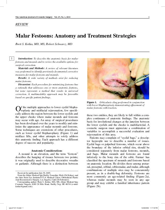
/
The American Journal of Cosmetic Surgery Vol. 27, No. 1, 2010 To descr The American Journal of Cosmetic Surgery Vol. 27, No. 1, 2010 To descr

The American Journal of Cosmetic Surgery Vol. 27, No. 1, 2010 To descr - PDF document

cheryl-pisano
cheryl-pisano . @cheryl-pisano
Follow
435 views
Uploaded On 2016-03-25

The American Journal of Cosmetic Surgery Vol. 27, No. 1, 2010 To descr - PPT Presentation

Received for publication July 30 2009 From the Allure Medical Spa Shelby Township Mich Dr Koltus and the Jules Stein Eye InstituteUCLA Medical Center Orbitofacial Plastic And Reconstructive Su ID: 269094

Received for publication July

Share:

Link:

Embed:

Download Presentation from below link

Download Pdf The PPT/PDF document "The American Journal of Cosmetic Surgery..." is the property of its rightful owner. Permission is granted to download and print the materials on this web site for personal, non-commercial use only, and to display it on your personal computer provided you do not modify the materials and that you retain all copyright notices contained in the materials. By downloading content from our website, you accept the terms of this agreement.


Presentation Transcript

The American Journal of Cosmetic Surgery Vol. 27, No. 1, 2010 To describe the anatomic basis for malar festoons and mounds and to review the available options for surgical correction. A review of relevant literature was performed to identify previously documented corrective measures for malar festoons and mounds. Received for publication July 30, 2009. From the Allure Medical Spa Shelby Township, Mich (Dr Koltus), and the Jules Stein Eye Institute/UCLA Medical Center, Orbitofacial Plastic And Reconstructive Surgery, Los Angeles, Calif (Dr Schwarcz).Corresponding author: Brett S. Kotlus, MD, MS, Allure Medical Spa, Cosmetic Surgery, 8180 26 Mile Road, Shelby Twp, MI 48316 (e-mail: kotlus@gmail.com).Malar Festoons: Anatomy and Treatment StrategiesBrett S. Kotlus, MD, MS; Robert Schwarcz, MD Figure 1. with lower blepharoplasty demonstrating effacement of malar festoons with traction. 1 j 2/29/2009 1:17:18 PM 12/29/2009 1:17:18 PM The American Journal of Cosmetic Surgery Vol. 27, No. 1, 2010transition between the lower eyelid and the cheek, sometimes extending to the cheek itself. Laxity of skin and atrophy of underlying soft tissues in this area results in the appearance of a structural descent. The guration of this apparent downward movement uenced by underlying retaining ligaments, including the orbitomalar ligament and the zygomatic The orbitoma-lar ligament (also known as the orbital retaining ligament) attaches from the infraorbital rim to the dermis and functioning as a skeletal support structure of the lower eyelid. With age, this ligament tends to attenuate, allowing for orbicularis descent as orbital fat simultaneously protrudes. The zycomaticus ligaments, with skeletal origins adjacent to those of rmly adherent to the dermis. As the cephalad tissues ate, they may hang over the dense or festoon. This anatomic boundary may be clinically apparent after surgery or trauma to the lower eyelids as ecchymosis is often distinctly limited by these Separation of the orbital septum from the capsulo-palpebral fascia with concurrent orbicularis oculi descent and prolapse of orbital fat can result in the outward pouching of tissues above the malar region,contributing to the downward forces inß uencing malar effect. At the same time, generalized atrophy of peri-orbital and malar fat can accentuate the loss of youth-ful contour where the lower eyelid blends with the ated, sagging appearance. The loss of contain several possible tissue layers. The adipose accumulations in the malar region consist of subcuta-neous fat, suborbicularis oculi fat, and preperiosteal fat. Hypertrophic and/or lax orbicularis oculi muscle can also contribute to a soft-tissue bulge in this area.(Figure 3). Excessive accumulations of malar ß uid can Figure 2. Festoons in a 73-year-old man with accompanying facial aging, and malar mounds in a 36-year-old woman. Figure 3. Malar festoon in a patient with unilateral facial jcs-27-01-10.indd 2 2/29/2009 1:17:21 PM 12/29/2009 1:17:21 PM The American Journal of Cosmetic Surgery Vol. 27, No. 1, 2010 result in a sponge effect, exacerbated by factors that uence systemic edematous states, such as increased sodium intake. Edematous festoons have also been described as a drug-associated side effect. Addition-ally, orbicularis oculi tonicity may facilitate lymphatic ow, and loss of muscle tone in this area can result in edematous festoons. This transient phenomenon has been seen after the injection of botulinum toxin type Iatrogenic orbicularis oculi denervation can likewise occur after surgical procedures that violate the pathway of the facial nerve Skeletal remodeling exerts a deeper structural inß u-The bony volume loss seen with aging leads to dimin-ishing malar projection and loss of skeletal support for the overlying soft tissues. Soft-tissue sagging and descent therefore ensue when skeletal reduction malar mounds and festoons and details of corrective ed from the OVID Medline primary author using a standardized form. The data abstraction form included Þ elds for year of publica-tion, method of festoon correction, sample size, and cation of 16 1906 to 2006 (one article was a republication). All articles described a surgical correction for mounds and/or festoons. Those including case series were presented in a retrospective manner. The most com-mon complications resulting from surgical corrections bulges. These data are summarized in the Table. Surgical Approaches described the direct excision of malar festoons in 1907: ÒTo excise the fold well away from the free margin of the lid, the fold is picked up between the thumb and index Þ nger of one hand and is trimmed away with sharp scissors. If the patient is Surgical Approaches to Malar Mound and Festoons jcs-27-01-10.indd 3 2/29/2009 1:17:22 PM 12/29/2009 1:17:22 PM The American Journal of Cosmetic Surgery Vol. 27, No. 1, 2010 eshy, the fatty tissue is freely trimmed away. ne silk collodion.ÓThis technique includes an external inci-because of aesthetic considerations, but it is consid-the inability to read due to visual obstruction. This technique was later readdressed by Netscher and Another approach used the subciliary, transcutane-ous lower eyelid incision. Klatsky and Manson cation of the traditional lower eyelid blepharoplasty that involved the dissection of separate aps in order to separate-ly manage the components of what they termed Òorbicularis festoonsÓ and Òsecondary bags.Ó In their ap is undermined beyond the ap extends only to the rim. The orbicularis ß ap is tightened to create a sling, and skin is then redraped and trimmed. They recognized that lower eyelid ectropion is a risk of this procedure, particularly if there is a lack of lower eyelid support. Farrior and Kassir detailed a modiÞ ap extends below the infraorbital rim to the inferior extent of the malar deformity. In addition, the subcu-taneous and suborbicularis fat is partially excised, and of the infraorbital rim. Farrior and Kassir hypothe-sized that fat dissection in these layers results in a Òfavorable Þ brosisÓ that improves the edema some-Alternatively, a myocutaneous ß ap has been described unit over the zygoma through a subciliary incision. xes the ß ap to the periosium lateral to the orbital rim for support.Suspension of an attenuated or elongated orbitomalar forcing the lower eyelid, and this may directly address a fundamental underlying cause of involutional The importance of repositioning descended subor-bicularis oculi fat (SOOF) has been stressed by Hoe- as a means of restoring the youthful midface convexity and managing malar festoons. This layer of fat, located posteriorly to the orbicularis oculi muscle, is suspended from the arcus marginalis with multiple aps are then redraped in an upward direction, with subcutaneous defatting of the skin ß ap and lateral Many address the lower eyelid and midcheek as a part of total facial rejuvenation, as has been advocated with the composite face-lift. He suggests rather than a superolateral direction, thus avoiding the also strives to maintain the continuity between the tect the branches of the facial nerve found on their posterior surface. Using a subciliary incision, Ham-raÕs approach releases the arcus marginalis before ap, in a separate dissection from a deep plane face-lift.The so-called midface lift, as has been described using a multitude of techniques, is another way to restore the natural contours of the eyelid/cheek region by repositioning, elevating, or suspending the com-monly descended malar fat pad. Commonly, the malar fat pad is anchored to the temporalis fascia, providing a superotemporal lift. A subperiosteal dissection may allow for more profound midface repositioning, but it can result in prolonged postoperative edema.cheek fat atrophy is often improved with Þ lling tech-niques. These attempt to efface folds and depressions by restoring volume to the region. Volume restoration can be accomplished via transposition of orbital fat autologous fat pearls or aspirated fat graft- and injectable to name a few. In most cases, the addition of volume alone may provide support to adjacent areas and act to disguise structural descent. The gamut of Þ llers and implants available can augment the facial frame-work in any layer, including subperiosteal, supraperi-osteal, intramuscular, subcutaneous, and intradermal. The evolving techniques and technologies used for lling are frequently combined with other surgical Attempts have been made to modify the fatty com-ponent of malar bags with suction lipectomy. Rosen-berg described improvement of the Òsaddlebag deformityÓ with suctioning in the immediate subder-a solitary procedure. A compressive postoperative dressing is applied, and the expected soft tissue contraction is exploited to achieve a regional improve-ment in soft-tissue sagging. This approach does not 3 jcs-27-01-10.indd 4 2/29/2009 1:17:23 PM 12/29/2009 1:17:23 PM The American Journal of Cosmetic Surgery Vol. 27, No. 1, 2010 address muscular or ligamentous attenuation and is most likely to beneÞ t a subset of patients with primarily a fat and/or ß uid collection. Improvement in the appearance of malar bags has periorbital resurfacing. The tightening effect of the carbon dioxide laser, for example, can act to efface Important variables to consider include the depth and precision of the resurfacing modality and its compat-ibility with the skin type, texture, and pigmentation of the patient. A potential for scarring, ectropion, and pigmentary aberrations exist with these treatments. Methods of fractional resurfacing have emerged that ated with nonfractional devices. Nonablative radiofre-quency has also been used in this area with modest success and fewer of the risks associated with ablative therapy. However, these techniques do not reposition descended structures but act to stimulate contraction and collagen formation. It is unclear how they modify Malar mounds and festoons are often an elusive target of facial rejuvenation plans. Over the years, many approaches have been suggested. Nine approaches are c anatomic Þ ndings of each patient, and therein lies the success of the surgical 1. Direct excision2. Skin-muscle ß ap3. Extended skin-muscle ß ap4. Orbital fat reduction or repositioning5. SOOF lift6. Midface lift7. Volume replacement8. Suction lipectomy9. Skin resurfacingFor an anatomic Þ nding, some combination of the following (the approaches combined are listed Prolapsed orbital fat (treatment approaches 2, 3, 4, Malar fat atrophy or descent (treatment approaches SOOF atrophy/descent (treatment approaches 3, 4, Bony loss (treatment approach 7)Clearly, there is no single procedure that remedies all varieties of mounds and festoons. Recurrence may be seen even after the most diligent surgical correc-tion, which can be frustrating for surgeons and patients. In our experience, patients with a ß at malar eminence or lack of youthful malar convexity are anatomically predisposed to prolonged postoperative A thorough examination with attention to periocular and midface Þ ndings is essential. The clinician must of dry eyes, BellÕs phenomenon, normal blink mecha-nism, and eyelid malposition. The degree of lower eyelid laxity and presence of canthal dystopia should or repositioning is required. Each of the following quality, dermatochalasis, steatoblepharon, orbicularis laxity and/or hypertrophy, descent of the malar fat pad, regional soft-tissue atrophy, skeletal architecture, degree of globe prominence, and localized aggrega-tions of fat and/or ß uid. The exact locations of each seated position. Intimate knowledge of these elements and festoons. An understanding of the various ana- ciency are cial in achieving satisfactory results. Ultimately, a multifaceted approach may be the best one, that is, concurrent extended lower blepharoplasty with lateral canthoplasty and fractional skin resurfacing. We will nd that there are more corrective options for malar mounds and festoons as we gain technological strides and further our understanding of this complex facial subunit.References 1. Furnas DW. Festoons, mounds, and bags of the eyelids and cheek. Clin Plast Surg 2. Kikkawa DO, Lemke BN, Dortzbach RK. cial musculoaponeurotic system ajcs-27-01-10.indd 5 2/29/2009 1:17:23 PM 12/29/2009 1:17:23 PM The American Journal of Cosmetic Surgery Vol. 27, No. 1, 2010 Ophthal Plast Reconstr Surg 3. Mendelson BC, Muzaffar AR, Adams WP. Sur-Plast Reconstr Surg 4. Hoenig JA, Shorr N, Goldberg RA. The versatile SOOF lift in oculoplastic surgery. Facial Plast Surg Clin North Am 5. Putterman, AM. Baggy eyelids have a single Plast Reconstr Surg 6. Butterwick KJ, Lack EA. Facial volume restora-tion with the fat autograft muscle injection technique. Dermatol Surg 7. Goldberg RA, McCann JD, Fiaschetti D, Ben Simon GJ. What causes eyelid bags? Analysis of 114 consecutive patients. Plast Reconstr Surg 8. Zide BM. Surgical Anatomy Around the Orbit: The System of Zones. Philadelphia, Pa: Lippincott Williams and Wilkins; 2005. 9. Webster PJ, Wulc AE, Moody BR, Dryden RM, Foster JA. Electrosurgical modiÞ cation of orbicularis oculi hypertrophy. Ophthal Plast Reconstr Surg10. Esmaeli B, Prieto VG, Butler CE, et al. Severe periorbital edema secondary to STI571 (Gleevec). 11. Goldman MP. Festoon formation after infraor-bital botulinum A toxin: a case report. Dermatol Surg12. Shaw RB, Kahn DM. Aging of the midface bony study. Plast Reconstr Surg13. Miller CC. The excision of bag-like folds of skin from the region about the eyes. 14. Netscher DT, Peltier M. Ancillary direct exci-sions in the periorbital and nasolabial regions for facial rejuvenation revisited. Aesthetic Plast Surg15. Klatsky SA, Manson PN. Separate skin and aps in lower-lid blepharoplasty. Reconstr Surg16. Farrior RT, Kassir RR. Management of malar folds in blepharoplasty. Laryngoscope17. Small RG. Extended lower eyelid blepharo-plasty. Arch Ophthalmol18. Kahana A, Lucarelli MJ. Adjunctive transcan-thotomy lateral suborbicularis fat lift and orbitomalar ligament resuspension in lower eyelid ectropion repair. Ophthal Plast Reconstr Surg19. Hamra ST. The zygorbicular dissection in composite rhytidectomy: an ideal midface plane. Reconstr Surg20. Hamra ST. Composite rhytidectomy. Reconstr Surg21. Anastassov GE, St Hillaire H. Periorbital and midfacial rejuvenation via blepharoplasty and sub-periosteal midface rhytidectomy. Int J Oral Maxillofac Surg22. Goldberg RA, Edelstein C. Shorr N. Fat reposi-tioning in lower blepharoplasty to maintain infraorbital rim contour. Facial Plast Surg23. Seiff SR. The fat pearl in ophthalmic plastic surgery: everyone wants to be a donor! 24. Kranendonk S, Obagi S. Autologous fat transfer for periorbital rejuvenation: indications, technique, and Dermatol Surg25. Carruthers JD, Carruthers A. Facial sculpting Dermatol Surg26. Flowers RS. Tear trough implants for correction of tear trough deformity. Clin Plast Surg27. Rosenberg GJ. Correction of saddlebag defor-mity of the lower eyelids by superÞ cial suction lipectomy. Plast Reconstr Surg28. Roberts TL. Laser blepharoplasty and laser resurfacing of the periorbital area. Clin Plast Surg29. Glavas IP, Purewal BK. Noninvasive techniques in periorbital rejuvenation. Facial Plast Surg30. Ruiz-Esparza J. Noninvasive lower eyelid blepharoplasty: a new technique using nonablative radiofrequency on periorbital skin. Dermatol Surg 6 jcs-27-01-10.indd 6 2/29/2009 1:17:23 PM 12/29/2009 1:17:23 PM Author: This article has been lightly edited for grammar, style, and usage. Please compare it with your original document and make changes on these pages. Please limit your corrections to substantive changes that affect meaning. If no change is required in response to a question, please write ÒOK as setÓ in the margin. Copy Author: Please check the date for Miller in paragraph 1 of ÒSurgical Approaches.Ó The text says 1907 and the reference says 1906. Copy editorAuthor: In paragraph 3 of ÒSurgical Approaches,Ó please add the manufacturerÕs name and location for the brand name Prolene. Copy editor4. Author: Please add the ending page number for reference 5. Copy editor5. Author: Please add the ending page number for reference 13. Copy editor6. Author: Reference 30 does not appear to be cited in text. Please cite or delete. Copy editor ajcs-27-01-10.indd 7 2/29/2009 1:17:23 PM 12/29/2009 1:17:23 PM