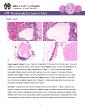
/
Figure Legend  Figure 1 Ovary  Cystin a female B6C3F1N mouse from a Figure Legend  Figure 1 Ovary  Cystin a female B6C3F1N mouse from a

Figure Legend Figure 1 Ovary Cystin a female B6C3F1N mouse from a - PDF document

claire
claire . @claire
Follow
342 views
Uploaded On 2022-08-26

Figure Legend Figure 1 Ovary Cystin a female B6C3F1N mouse from a - PPT Presentation

Ovary 150 Cyst 1 Ovary 150 Cyst cysts increase with age and the walls become so thin that identifying features are lost The pathogenesis of ovarian cysts is often unknown however in mice s ID: 942044

figure ovary mouse cyst ovary figure cyst mouse female cysts eds study origin chronic cystin b6c3f1 toxicity ovarian follicular

Share:

Link:

Embed:

Download Presentation from below link

Download Pdf The PPT/PDF document "Figure Legend Figure 1 Ovary Cystin a ..." is the property of its rightful owner. Permission is granted to download and print the materials on this web site for personal, non-commercial use only, and to display it on your personal computer provided you do not modify the materials and that you retain all copyright notices contained in the materials. By downloading content from our website, you accept the terms of this agreement.


Presentation Transcript

Ovary – Cyst Figure Legend: Figure 1 Ovary - Cystin a female B6C3F1/N mouse from a chronic study. The cyst is adjacent to a hematocyst(arrow), which contains blood.Figure 2Ovary - Cystin a female B6C3F1/N mouse from a chronic study (higher magnification of Figure 1). The cyst contains pale acellular eosinophilic follicular fluid and a thin outer wall lined by one or two layers of cuboidal or flattened granulosa cells. Figure 3 Ovary - Cystin a female B6C3F1/N mouse from a chronic study. The large cyst is the predominating feature of this ovary. Figure 4 Ovary - Cystin a female B6C3F1/N mouse from a chronic study (higher magnification of Figure 3). Flattened epithelium lines the cyst wall, flocculent eosinophilic material is present in the lumen.Comment: Ovarian cysts are a common finding in rats and mice(Figure 1, Figure 2,Figure 3,Figure 4). Ovarian cysts may be unilateral or bilateral, single or multiple, and may becomequite large. Ideally, cysts should be classified by origin, location, and/or morphologic appearance (follicular, luteal, epithelial, rete ovarii, bursal, paraovarian), whichmay provide information as to

pathogenesis in toxicity or carcinogenicity studies. However, origin may be difficult to assign because the size and number of 1 Ovary – Cyst cysts increase with age, and the walls become so thin that identifying features are lost. The pathogenesis of ovarian cysts is often unknown; however, in mice some of them may be omesonephric duct origin.Recommendation: Ovary - Cyst should be diagnosed but not assigned a severity grade. Whenever possible, the type of cyst (follicular, luteal, epithelial, rete ovarii, bursal, paraovarian) should be included in the diagnosis as a modifier. The diagnosis of Ovary - Cyst, with no modifier, is reserved for those instances when the origin of the cyst cannot be determined.References:Alison RH, Morgan KT, Montgomery CA. Ovary. In: Pathology of the Fischer Rat: Reference and Atlas(Boorman GA, Eustis SL, Elwell MR, Montgomery CA, MacKenzie WF, eds). Academic PressSan Diego, CA, Davis BJ, Dixon D, Herbert RA.1999.Ovary, viduct, terus, ervix agina. In: Pathology of the Mouse: Reference and Atlas(Maronpot RR, BoormanGA, Gaul BW, eds). Cache River PressVienna, IL, 444.Dixon D, Heider K, E

lwell MR.Incidence of onneoplastic esions in istorical ontrol ale and emale Fischer344 ats from 90-day oxicity tudies. Toxicol Pathol23:338Abstract: http://www.ncbi.nlm.nih.gov/pubmed/7659956 Faccini JM, Abbott DP, Paulus GJJ. 1990. Female enital ract. In: Mouse Histopathology: A Glossary for Use in Toxicity and Carcinogenicity Studies.Elsevier,Amsterdam,Greaves P.Female enital ract. In: Histopathology of Preclinical Toxicity Studies: Interpretation and Relevance in Drug Safety Evaluation, 4ed. ElsevierAmsterdam, 667Long G. 2002. Apparent esonephric uct (ete nlage) rigin for ysts and roliferative pithelial esions in the mouse ovary. Toxicol Pathol 30:592Abstract: http://www.ncbi.nlm.nih.gov/pubmed/12371668 Maekawa A, Maita K, Harleman JH.1996.Changes in the vary. In:Pathobiology of the Aging MouseVol 1 (Mohr U, Dungworth DL, Capen CC, Carlton WW, Sundberg JP, Ward JM, eds).ILSI PressWashington, DC, 451467.Maekawa A, Yoshida A.Susceptibility of the emale enital ystem to oxic ubstances. In:Pathobiology of the Aging Mouse, Vol 1 (Mohr U, Dungworth DL, Capen CC, Carlton WW, Sundberg JP, Ward JM, eds). ILSI Press, Washington, DC, 2