/
Respiratory Emergencies Respiratory Emergencies

Respiratory Emergencies - PowerPoint Presentation

conchita-marotz
conchita-marotz . @conchita-marotz
Follow
517 views
Uploaded On 2017-04-07

Respiratory Emergencies - PPT Presentation

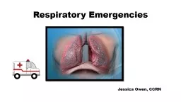
Erin Moorcones RN MSN CPNP Anatomy and Physiology Anatomy Adequate amount of O2 delivered to cell The affinity of hemoglobin for oxygen The ease in which hemoglobin release oxygen to cell ID: 534800

airway pulmonary cough chest pulmonary airway chest cough oxygen asthma dyspnea pneumonia death hemoglobin pain severe edema children exam

Share:

Link:

Embed:

Download Presentation from below link

Download Presentation The PPT/PDF document "Respiratory Emergencies" is the property of its rightful owner. Permission is granted to download and print the materials on this web site for personal, non-commercial use only, and to display it on your personal computer provided you do not modify the materials and that you retain all copyright notices contained in the materials. By downloading content from our website, you accept the terms of this agreement.


Presentation Transcript

Slide1

Respiratory Emergencies

Erin

Moorcones

, RN, MSN, C-PNPSlide2

Anatomy and PhysiologySlide3
Slide4

AnatomySlide5

Adequate amount of O2 delivered to cell

The affinity of hemoglobin for oxygen

The ease in which hemoglobin release oxygen to cell

Cellular oxygenationSlide6

Affinity of

Hgb

for oxygen is described in the oxygen-hemoglobin dissociation curve.

If it shifts to left-

hgb

picks up o2 easier in the lungs, but does not release to the tissues as easy.

When it shifts to right,

oxygen

uptake by

hgb

is slower, but it is transported to cells easier.This is affected by temp, acid-base balance, and co2.Slide7

Normal gas exchangeSlide8

Patient AssessmentSlide9

Airway First!Work of breathing, nasal flaring, retractions, accessory muscle use

Occupational exposure

Smoking history

AssessmentSlide10

Usually caused by a virus- Influenza A &B,

parainfluenza

, RSV, rhinovirus,

coxsackie

virus, and adenovirus.

Secondary infections possible from

mycoplasma

pneumoniae

,

haemophilus

influenzae.Most common in smokers, young children, and during winter months.

Acute bronchitisSlide11

S/S- sore throat, stuffy nose and dry non productive cough (may be worse at night). Sometimes with low grade fever, chest discomfort and fatigue.

Aggravating factors- cold, talking, deep breathing, laughing

Diagnosis- clinical exam.

Would you get an

xray

? Why?

Treatment- Fluids, avoid irritants and smoking. ABX?

Acute bronchitisSlide12

Causes- bacterial, viral, or fungal

Primarily effects young children, debilitated adults, those with underlying chronic conditions.

6

th

leading cause of death in US, and leading cause of death in the elderly.

What would be RF for pneumonia?

PneumoniaSlide13

S/S- fever, malaise, cough, hemoptysis, dyspnea,

pleuritic

chest symptoms.

Physical exam- crackles on auscultation that don’t clear with coughing, diminished breath sounds/cough, fever.

Alternate s/s- vomiting, abdominal pain, mental status changes, headaches.

Diagnosis-

xray

, CBC, pulse ox,

abg

Core Measures…antibiotics within 4 hours!

PneumoniaSlide14
Slide15
Slide16
Slide17

One of joint Commissions recommendations

Those

pts

presenting to ED must have O2 assessment or ABG

During triage patient’s pneumococcal vaccination status and smoking

hx

must be documented (cessation material must be provided to ay admitted smokers).

BC must be done prior to any

abx

administration.

ABX should be administered w/I 4 hours of arrival to ER

Core Measures-

Community Acquired PneumoniaSlide18

Obstructive disease caused by airway inflammation and

hyperreactivity

.

Males more affected than females.

30% of those diagnosed with asthma as children will have it as adults.

+ family

hx

in more than 1/3

AsthmaSlide19
Slide20
Slide21

History- patterns, triggers, allergies

Physical exam- rhinitis, sinusitis, or nasal polyps, wheezes, and prolonged expiratory phase.

Labs- CBC with diff, nasal smears, and sputum specimen.

In those older then 5-

spirometry

, peak expiratory flow rate

Asthma DiagnosisSlide22

Goal is to relieve hypoxemia and airflow obstruction and decreasing airway inflammation

O2, SABA’s, systemic steroids

Asthma TreatmentSlide23

Emphysema & Bronchitis Slide24

650, 000 patients diagnosed annually

10% of patient with fatal PE die within 1 hour of onset of symptoms.

90% of thrombi originate in deep vessels of the leg.

Stasis of blood damage to epithelium of vessel wall, and alterations in coagulation ( Virchow’s triad)- lead to thrombi formation.

Thrombi becomes dislodged and travels and lodges in pulmonary vessel, obstructing blood flow.

Pulmonary EmbolusSlide25

s/s- dyspnea, tachycardia, tachypnea, restlessness, anxious, severe chest pain, cough, fever

If large vessel occluded can cause hypotension and right sided heart failure.

Pulmonary EmbolusSlide26
Slide27

Diagnosis and treatment

DX- identify VQ mismatch, ABG, 12 lead EKG, CXR, labs, CT

TX- ABC’s, pain meds, heparin protocols, possible surgical intervention

Pulmonary EmbolusSlide28

Cardiogenic vs

Non cardiogenic

Pulmonary EdemaSlide29
Slide30

Severe dyspnea, diaphoresis, hypertension, tachycardia, anxiety, tachypnea.

Pitting edema, weight gain, bounding pulse, skin cool/pale, cough with frothy white sputum, cyanosis

S/S of pulmonary edemaSlide31

Goal- increase oxygenation, decrease cardiac workload, increase cardiac function

Bronchodilators- decreased bronchospasm

Digoxin- increase contractility, decrease HR

Dobutamine

-

inc

contractility, decrease PVR

Diuretic therapy

Positioning

Treatment of pulmonary edemaSlide32

What is this?Slide33

Usually occur in males 20-40y/o, tall and thin

Spontaneous- usually caused by ruptured emphysematous bleb.

Smokers increased risk

Iatrogenic causes- trauma, insertion SVC, mechanical ventilation

PneumothoraxSlide34

S/S- chest pain on affected side, sob

Larger

pneumo

-

subQ

emphysema, cyanosis, severe sob, cyanosis

Treatment- needle aspiration or chest tube for larger

pneumo

, if <15% you can observe

PneumothoraxSlide35

Hemoglobin has greater affinity for CO than for oxygen, resulting in O2 being displaced by CO.

Carboxyhemoglobin

(

COHb

) levels >10% indicate CO exposure

Smokers of individual exposed to automobile exhaust can have baseline 10-15%

5-10% experience HA or vertigo

10-20% experience HA, nausea, vomiting, loss of coordination, dyspnea,

pt

may be flushed

20-30%- confused, lethargic, visual disturbance

Carbon Monoxide PoisoningSlide36

ST depression due to myocardial hypoxia

COHb

levels 40-60%- comatose, seizures,

ectopy

COHB >60% incompatible with life

Pulse ox not reliable indicator of oxygenation status

Treatment- if cyanide suspected, treat with antidote

CO poisoningSlide37

Most common in children and older adults

Upper airway presentation- severe distress-no distress, can present like choking patient.

Lower airway- can be asymptomatic

Treatment- BLS, visualization with laryngoscope, lower airway requires bronchoscopy

Foreign Body AspirationSlide38
Slide39

5

th

leading cause of accidental death in US

Approx

8000 deaths/

yr

Hypoxia is cause of death in all drowning patients

Wet drowning- asphyxiation results in relaxation of airways, allowing H2O to enter airway

Dry drowning- 10-20%, aspiration of H2O does not occur because airway doesn’t relax until cardiac arrest

What is significant info to obtain is submersion injuries?

Submersion InjuriesSlide40

Assessing ventilation status

Address hypothermia

Fluid resuscitation

CXR

Gastric tube insertion

Treating submersion injuriesSlide41