/
Reticular fatty degeneration Reticular fatty degeneration

Reticular fatty degeneration - PowerPoint Presentation

conchita-marotz
conchita-marotz . @conchita-marotz
Follow
421 views
Uploaded On 2017-05-03

Reticular fatty degeneration - PPT Presentation

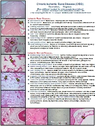
Poorly forming new bone Ischemic Bone Diseases Avascular necrosis Dead bone large regions with missing osteocytes Osteonecrosis Dead bone all osteocytes are missing Also used as umbrella term for ID: 544170

ischemic bone cells marrow bone ischemic marrow cells fibrous focal myelofibrosis tissue fat osteocytes missing chronic scar thin osteosclerosis trabeculae intramedullary variant

Share:

Link:

Embed:

Download Presentation from below link

Download Presentation The PPT/PDF document "Reticular fatty degeneration" is the property of its rightful owner. Permission is granted to download and print the materials on this web site for personal, non-commercial use only, and to display it on your personal computer provided you do not modify the materials and that you retain all copyright notices contained in the materials. By downloading content from our website, you accept the terms of this agreement.


Presentation Transcript

Slide1

Reticular fatty degeneration

Poorly forming

new bone

Ischemic Bone Diseases:

Avascular necrosis

Dead bone; large regions with missing osteocytes.

Osteonecrosis:

Dead bone (all osteocytes are missing). Also used as umbrella term for all ischemic bone disease. Partially nonviable bone: Ischemically damaged bone shows clusters (not scattered) of missing or pyknotic osteocytes (must process bone properly to evaluate). Ischemic osteosclerosis: Sheets of lamellar bone and/or thickened trabeculae, usually with focal regions showing missing osteocytes; often with microcracks. Bone scar: Variant of ischemic osteosclerosis (see above); atypical healing from medullary infarction. Condensing osteitis: Variant of ischemic osteosclerosis (see above); at apex of nonviable tooth; response to dental infection. Honeycombed bone: Same as ischemic marrow atrophy (see below). Poorly forming new bone: Region of immature, newly forming bone (lamellar, typically) which did not remodel to full maturity; no remaining osteoblastic activity; fibrous background changes to mature fat cells.Ischemic Marrow Diseases: Marrow congestion (dilated vessels): Normal marrow except that capillaries or veins are dilated more than 6 RBC diameters; may be cause of pain. Ischemic myelofibrosis: Fat cells slowly are replaced by a loose, wispy fibrous tissue which seems to be streaming between the fat cells; a few mast cells, perhaps a few chronic inflammatory cells; often multifocal. Reticular fatty degeneration: Mild version of ischemic myelofibrosis; fibers look ragged. Intramedullary fibrous scar: Dense collagen fibers aggregated into scar tissue with very little vascularity or inflammation; sharp outer borders; may be myxoid in areas. Bone marrow edema: Classic CIBD, with dilated vessels, myelofibrosis, plasmostasis, focal hemorrhage, focal fat necrosis (all types), a few mast cells, a few chronic inflammatory cells; involved bone is almost always viable. Regional ischemic osteoporosis: Low bone density with thin, widely spaced trabeculae, a thin cortex, and diffuse or multifocal signs of ischemic marrow damage. Focal osteoporotic marrow defect: Low bone density with thin, widely spaced, inactive trabecula, a thin cortex, and minimal, if any, signs of ischemic marrow damage. Ischemic marrow atrophy (honeycombed bone): Inactive, almost always viable bony trabeculae with no or almost no attached fatty marrow or hematopoietic marrow; may have small amounts of attached fibrous tissue; only a valid feature if no rotary instrument touched the cancellous bone sample. Ischemic cavitation: Intramedullary void, greater than 5 mm. diameter, with little or no soft tissue lining; scraping walls may yield: fibrous tissue, viable trabeculae, scattered chronic Inflammatory cells, plasmostasis, focal hemorrhage, ischemic myelofibrosis; surgeon must indicated the presence of the void. Ischemic microcavitations are small variants. Traumatic bone cyst: Variant of ischemic cavitation (see above), found primarily in young persons, usually asymptomatic, easily treated with curettage (inducing new hemorrhage).

Avascular necrosis

Chronic Ischemic Bone Disease (CIBD)

Descriptions

,

Diagnoses

[See additional handout for microscopic descriptions]

Dr. J.E. Bouquot, University of Texas Dental Branch, Houston, Texas

Jerry.Bouquot@uth.tmc.edu; 713-500-4420; additional photos: maxillofacialcenter.com

Honeycombed bone

Ischemic osteosclerosis

Focal osteoporotic marrow defect

Bone marrow edema

Intramedullary fibrous scar

Ischemic cavitation

Ischemic myelofibrosis