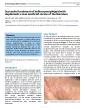
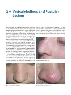
/
Vesiculobullous and Pustular LesionsWhile	vesiculous,	bullous	and	pust Vesiculobullous and Pustular LesionsWhile	vesiculous,	bullous	and	pust

Vesiculobullous and Pustular LesionsWhile vesiculous, bullous and pust - PDF document

conchita-marotz
conchita-marotz . @conchita-marotz
Follow
370 views
Uploaded On 2015-10-02

Vesiculobullous and Pustular LesionsWhile vesiculous, bullous and pust - PPT Presentation

Fig21Regional hyperhidrosis Fig23 Nose The Importance from a Dermatological Point of View and the face so these pruritic lesions can be prominent on the nose Fig Ophthalmic zoster resulti ID: 147586

Fig.2.1.Regional hyperhidrosis Fig.2.3. Nose. The Importance from

Share:

Link:

Embed:

Download Presentation from below link

Download Pdf The PPT/PDF document "Vesiculobullous and Pustular LesionsWhil..." is the property of its rightful owner. Permission is granted to download and print the materials on this web site for personal, non-commercial use only, and to display it on your personal computer provided you do not modify the materials and that you retain all copyright notices contained in the materials. By downloading content from our website, you accept the terms of this agreement.


Presentation Transcript

Vesiculobullous and Pustular LesionsWhile vesiculous, bullous and pustular elementary lesions can be seen in different diseases, vesicles and bullae (blisters) can Fig.2.1.Regional hyperhidrosis Fig.2.3. Nose. The Importance from a Dermatological Point of View and the face, so these pruritic lesions can be prominent on the nose (Fig.). Ophthalmic zoster, resulting from the reactivation of Varicella zoster virus, involves the �rst branch of the trigeminal nerve causing unilateral vesicles on the eyelid, forehead and in front of the scalp. Ocular involvement should be suspected when blisters are seen unilaterally on the nose, indicating involvement of the nasocilliary branch (Hutchinson sign) (Fig.). Afterwards, vesicles become purulant and develop into necrotic crusts and ulcers. Patients usually suffer from intense pain. Lesions may last longer when compared with herpes zoster lesions of the trunk. Systemic antiviral therapy is indicated to prevent the complications like uveitis and keratitis. Post-herpetic neuralgia is a frequent problem in patients with ophthalmic zoster.Impetigo, a super�cial bacterial infection, develops on the nose usually after long lasting �u infection. It is most common among children (Fig.). Clinically it can be seen as bullous and nonbullous impetigo (impetigo contagiosa). Staphylococcus aureus is responsible for the bullous form and Streptococcus sp. for the nonbullous form, however this distinction is currently not considered important. As S. aureus colonizes the nasal mucosa, the infection may start in the perinasal area in carriers of the bacteria and spread to the periphery. Blisters typically rupture and leave characteristic honey-coloured crusts. Perinasal crusting and serum leakage are commonly observed presentations (Fig.). The infection responds well to antibiotic therapy and all lesions heal without scarring. Staphylococcal folliculitis is usually seen Fig.2.7.ImpetigoFig.2.6.Impetigo Fig.2.5.Ophtalmic zoster Fig.2.4.Varicella Vesiculobullous and Pustular Lesions on the ala nasi as super�cial pustules (Fig.). Another staphylococcal infection, furuncle, presents with a deeply located in�ammatory nodule with a central pustule (Fig.). Although low, there is a risk of development of cavernous sinus thrombosis after pyodermas on the nose, therefore treatment with systemic antibiotics is necessary.Cat scratch disease, which starts as a papule or a crusted pustule at the site of inoculation after being scratched by a cat, is a chronic infection caused by a gram negative bacillus, called Bartonella henselae. Abscess like lesions usually involving the tip of the nose, can be observed. It is one of the most common causes of lymphadenopathy lasting more than 3-4 weeks, in childhood and puberty. This infection usually regresses spontaneously. Acne vulgaris is commonly seen on the nose where the hair follicles are dense and the secretion of seborrheic glands is increased. Lesions also involve other parts of the face and sometimes the ears. In�ammatory papules, pustules, cysts and comedones can frequently be observed together (Fig.). Some pustules may impetiginize and be crusted (Fig.), and cystic lesions may rarely develop into abscesses. Demodex folliculorum mites are located in the hair follicles, and under some conditions they may be pathogenic causing demodicosis. Nose is one of the typical sites of demodicosis because of excessive production of sebum which is used as food by the mites. A like appearance with follicular papules and pustules are observed, especially when the immune system is suppressed. Epidermal growth factor receptor (EGFR) Fig.2.8.Staphyloccal folliculitis Fig.2.9.Furuncle Fig.2.10.Acne vulgaris Fig.2.11.Acne vulgaris. Secondary impetiginization Nose. The Importance from a Dermatological Point of View antogonists-induced drug eruption may present with pustules Pemphigus vulgaris should be suspected when there are non-healing erosions and crusts on the nose (Fig.). Intact �accid blisters are not common in this area. These erosions are usually located on the ala nasi uni- or bilaterally and can even cover the entire nose. Although rare, it must be remembered that pemphigus vulgaris may be restricted to the nose and may relapse after therapy at the same site. In some cases pemphigus vulgaris may involve the nasal mucosa causing chronic erosions, crusting and sometimes nasal congestion, discharge or epistaxis. Bullous pemphigoid may occasionally be seen on the nose both with intact bullae and erosions. Linear IgA dermatosis may present with tense nasal bullae on an erythematous base in adults. Similar lesions may be seen in chronic bullous dermatosis of childhood around the ages of 4 and 5. Bullae on other parts of the body mostly accompany nasal lesions in both diseases. Patients may complain of pruritus and burning sensation. Erosive and crusted lesions may be encountered on the cheeks, eyelids and nose in patients with Stevens-Johnson syndrome, a severe form of erythema multiforme which is usually caused by medications. Oral mucosal involvement with crusting on the lips is Hydroa vacciniforme is a type of photodermatosis seen in childhood. It causes recurrent crops of umbilicated, tiny vesicles on the nose and cheeks which evolve into hemorrhagic, necrotic or crusted lesions. They resolve typically Fig.2.12.Epidermal growth factor receptor antogonists-induced drug eruption Fig.2.13.Pemphigus vulgaris Fig.2.14.Pemphigus vulgaris Fig.2.15.Hydroa vacciniforme Vesiculobullous and Pustular Lesions with variola-like scars (Fig.). Actinic prurigo is another photodermatosis affecting the face. It is more commonly seen in girls at 8 to 14 years of age. They may also have family history. Papules and vesicles which tend to eczematize over time are observed on sun-exposed areas like nose, cheeks, forehead and ears. Porphyria cutanea tarda, the most common form of porphyrias, involves sun-exposed areas causing bullae, erosions, crusts, dyspigmentation and atrophic scars on the nose (Fig.). Since acute photosensitivity is rarely seen, patients may deny the role of the sun. Milia, large comedones and hypertrichosis may also be observed on the face in the course of the disease. In congenital erythropoietic porphyria (Günther’s disease) there may be atrophic scars on the nose due to recurrent vesiculobullous lesions. These may be present since birth or develop secondary to infections. In addition, severe photosensitivity and growth retardation are seen in these children. The diagnostic clinical feature of the disease is erythrodontia of both the primary and permanent Fig.2.16.Porphyria cutanea tarda