/
GASTROSCHISIS DIAGNOSED AT 13 WEEKS GESTATION GASTROSCHISIS DIAGNOSED AT 13 WEEKS GESTATION

GASTROSCHISIS DIAGNOSED AT 13 WEEKS GESTATION - PDF document

cora
cora . @cora
Follow
343 views
Uploaded On 2022-09-23

GASTROSCHISIS DIAGNOSED AT 13 WEEKS GESTATION - PPT Presentation

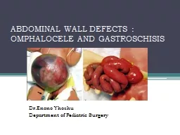
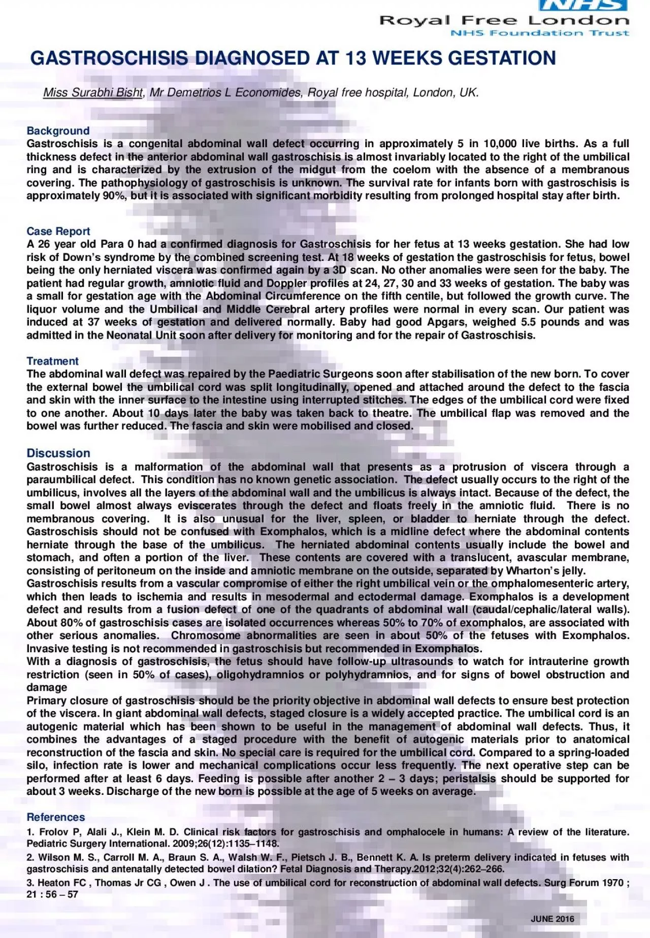
Miss Surabhi Bisht Mr Demetrios L Economides Royal free hospital London UK Background Gastroschisis is a congenital abdominal wall defect occurring in approximately 5 in 10 000 live births ID: 955867

abdominal gastroschisis wall defect gastroschisis abdominal defect wall umbilical weeks bowel gestation exomphalos cord defects baby results diagnosis fetus

Share:

Link:

Embed:

Download Presentation from below link

Download Pdf The PPT/PDF document "GASTROSCHISIS DIAGNOSED AT 13 WEEKS GEST..." is the property of its rightful owner. Permission is granted to download and print the materials on this web site for personal, non-commercial use only, and to display it on your personal computer provided you do not modify the materials and that you retain all copyright notices contained in the materials. By downloading content from our website, you accept the terms of this agreement.


Presentation Transcript

GASTROSCHISIS DIAGNOSED AT 13 WEEKS GESTATION Miss Surabhi Bisht , Mr Demetrios L Economides, Royal free hospital, London, UK . Background Gastroschisis is a congenital abdominal wall defect occurring in approximately 5 in 10 , 000 live births . As a full thickness defect in the anterior abdominal wall gastroschisis is almost invariably located to the right of the umbilical ring and is characterized by the extrusion of the midgut from the coelom with the absence of a membranous covering . The pathophysiology of gastroschisis is unknown . The survival rate for infants born with gastroschisis is approximately 90 % , but it is associated with significant morbidity resulting from prolonged hospital stay after birth . Discussion Gastroschisis is a malformation of the abdominal wall that presents as a protrusion of viscera through a paraumbilical defect . This condition has no known genetic association . The defect usually occurs to the right of the umbilicus, involves all the layers of the abdominal wall and the umbilicus is always intact . Because of the defect, the small bowel almost always eviscerates through the defect and floats freely in the amniotic fluid . There is no membranous covering . It is also unusual for the liver, spleen, or bladder to herniate through the defect . Gastroschisis should not be confused with Exomphalos, which is a midline defect where the abdominal contents herniate through the base of the umbilicus . The herniated abdominal contents usually include the bowel and stomach, and often a portion of the liver . These contents are covered with a translucent, avascular membrane, consisting of peritoneum on the inside and amniotic membrane on the outside, separated by Wharton’s jelly . Gastroschisis results from a vascular compromise of either the right umbilical vein or the omphalomesenteric artery, which then leads to ischemia and results in mesodermal and ectodermal damage . Exomphalos is a development defect and results from a fusion defect of one of the quadrants of abdominal wall (caudal/cephalic/lateral walls) . About 80 % of gastroschisis cases are isolated occurrences whereas 50 % to 70 % of exomphalos, are associated with other serious anomalies . Chromosome abnormalities are seen in about 50 % of the fetuses with Exomphalos . Invasive testing is not recommended in gastroschisis but recommended in Exomphalos . With a diagnosis of gastroschisis, the fetus should have follow - up ultrasounds to watch for intrauterine growth restriction (seen in 50 % of cases), oligohydramnios or polyhydramnios, and for signs of bowel obstruction and damage Primary closure of gastroschisis should be the priority objective in abdominal wall defects to ensure best protection of the viscera . In giant abdominal wall defects, staged closure is a widely accepted practice . The umbilical cord is an autogenic material which has been shown to be useful in the management of abdominal wall defects . Thus, it combines the advantages of a staged procedure with the benefit of autogenic materials prior to anatomical reconstruction of the fascia and skin . No special care is required for the umbilical cord . Compared to a spring - loaded silo, infection rate is lower and mechanical complications occur less frequently . The next operative step can be performed after at least 6 days . Feeding is possible after another 2 – 3 days ; peristalsis should be supported for about 3 weeks . Discharge of the new born is possible at the age of 5 weeks on average . References 1 . Frolov P, Alali J . , Klein M . D . Clinical risk factors for gastroschisis and omphalocele in humans : A review of the literature . Pediatric Surgery International . 2009 ; 26 ( 12 ) : 1135 – 1148 . 2 . Wilson M . S . , Carroll M . A . , Braun S . A . , Walsh W . F . , Pietsch J . B . , Bennett K . A . Is preterm delivery indicated in fetuses with gastroschisis and antenatally detected bowel dilation? Fetal Diagnosis and Therapy . 2012 ; 32 ( 4 ) : 262 – 266 . 3 . Heaton FC , Thomas Jr CG , Owen J . The use of umbilical cord for reconstruction of abdominal wall defects . Surg Forum 1970 ; 21 : 56 – 57 JUNE 2016 Case Report A 26 year old Para 0 had a confirmed diagnosis for Gastroschisis for her fetus at 13 weeks gestation . She had low risk of Down’s syndrome by the combined screening test . At 18 weeks of gestation the gastroschisis for fetus, bowel being the only herniated viscera was confirmed again by a 3 D scan . No other anomalies were seen for the baby . The patient had regular growth, amniotic fluid and Doppler profiles at 24 , 27 , 30 and 33 weeks of gestation . The baby was a small for gestation age with the Abdominal Circumference on the fifth centile, but followed the growth curve . The liquor volume and the Umbilical and Middle Cerebral artery profiles were normal in every scan . Our patient was induced at 37 weeks of gestation and delivered normally . Baby had good Apgars , weighed 5 . 5 pounds and was admitted in the Neonatal Unit soon after delivery for monitoring and for the repair of Gastroschisis . Treatment The abdominal wall defect was repaired by the Paediatric Surgeons soon after stabilisation of the new born . To cover the external bowel the umbilical cord was split longitudinally, opened and attached around the defect to the fascia and skin with the inner surface to the intestine using interrupted stitches . The edges of the umbilical cord were fixed to one another . About 10 days later the baby was taken back to theatre . The umbilical flap was removed and the bowel was further reduced . The fascia and skin were mobilised and closed .