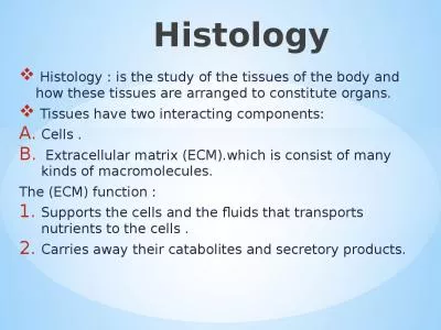
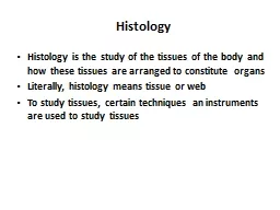
/
 Histology Histology is the study of the tissues of the body and how these tissues are  Histology Histology is the study of the tissues of the body and how these tissues are

Histology Histology is the study of the tissues of the body and how these tissues are - PowerPoint Presentation

danika-pritchard
danika-pritchard . @danika-pritchard
Follow
347 views
Uploaded On 2020-04-02

Histology Histology is the study of the tissues of the body and how these tissues are - PPT Presentation

organs Literally histology means tissue or web To study tissues certain techniques an instruments are used to study tissues Tissue Preparation For conventional bright field light microscopy we follow the following steps ID: 774747

tissue microscopy components dyes tissue microscopy components dyes staining microscope stain charged field embedding tissues study cutting solution positively

Share:

Link:

Embed:

Download Presentation from below link

Download Presentation The PPT/PDF document " Histology Histology is the study of the..." is the property of its rightful owner. Permission is granted to download and print the materials on this web site for personal, non-commercial use only, and to display it on your personal computer provided you do not modify the materials and that you retain all copyright notices contained in the materials. By downloading content from our website, you accept the terms of this agreement.


Presentation Transcript

Slide1

Histology

Histology is the study of the tissues of the body and how these tissues are arranged to constitute

organs

Literally, histology means tissue or web

To study tissues, certain techniques an instruments are used to study tissues

Slide2

Tissue Preparation

For conventional bright field light microscopy, we follow the following steps:

Fixation:

0.5 X0.5 cm tissue is added to a small jar containing the fixative to:

Prevent autolysis

Terminate cell metabolism

Kill bacteria

Harden tissue

The fixative used is usually Formalin 10% solution

Slide3

Tissue Preparation

2. Dehydration:

Ascending grades of alcohol are used to take water out of the specimen.

3. Clearing:

To clear the sample using solution miscible in alcohol and embedding medium. Solution used is Xylene

Slide4

Tissue Preparation

4. Embedding: Putting the specimen in embedding medium (Paraffin) to enable us to cut the sections5. Cutting: Cutting thin sections (5µm) by a microtome using steel knife6. Mounting: Putting the section on a glass slide using adhesive material (Egg albumin)

Slide5

Staining the Slide

Sections must pass through many steps before they are stained and be visualized in the light microscope

Principles of staining:

Staining is to impart colors to different tissue components.

Staining is a reaction between stain and tissue component

Slide6

Staining the Slide

Stains are either positively or negatively charged

Tissue components also are negatively and positively charged

Basic dyes (

Hematoxylene

) are bound to negatively charged tissue components

Acidic dyes

(Eosin) are bound to positively charged tissue components

Slide7

Basic dyes stain:

Heterochromatin

Nucleic acids

Ribosomes

Cartilage

Acidic dyes stain:

Filaments

Mitochondria

Collagen

Muscle fibers

Slide8

Additional Dyes

Many tissue components can not be stained with (

Hematoxylene

and Eosin).

Other dyes are used to specifically stain certain tissue components

Resorcin-Fuchsin

for elastic fibers

Silver stain for reticular fibers and basement membrane

Periodic-Acid

Schiff (PAS) Reaction for CHO

Slide9

Visualizing Slides

Bright field microscope is used to visualize specimens on glass slidesThe resolution of the light microscope is 0.2µmResolution is defined as( The smallest distance between 2 points can be seen as 2 points)

Slide10

Resolution

Human EyeLight microscopeElectron microscopeAtomic force microscope

0.2mm

0.2µm

0.2nm

0.05nm

Slide11

Study Techniques

Microscopy

Bright Field Microscopy

Dark field Microscopy

Electron Microscopy (Transmission and Scanning)

Fluorescent microscopy

Confocal microscopy

Atomic force microscopy

Histochemistry

and

Cytochemistry

Immunocytochemistry and Hybridization

Autoradiography

Slide12

Fomalin

Ethyle alcoholXyleneParaffinMicrotomeSteel knifeGlass slideH&ELight microscope

GlutaraldehydeAcetonePropylene oxideEpoxy resinUltramicrotomeDiamond knifeMetal gridUranyl acetateElectron microscope

Fixation

Dehydration

Clearing

Embedding

Cutting

Knife

Mounting

Staining

Viewing

Slide13

a

Slide14

Slide15

Slide16

a

Slide17

a

Slide18