/
neuro-op 	 Howard R Krauss, MD neuro-op 	 Howard R Krauss, MD

neuro-op Howard R Krauss, MD - PowerPoint Presentation

danya
danya . @danya
Follow
342 views
Uploaded On 2022-06-14

neuro-op Howard R Krauss, MD - PPT Presentation

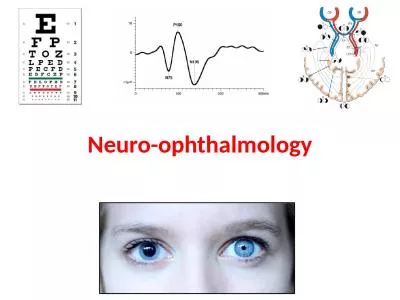
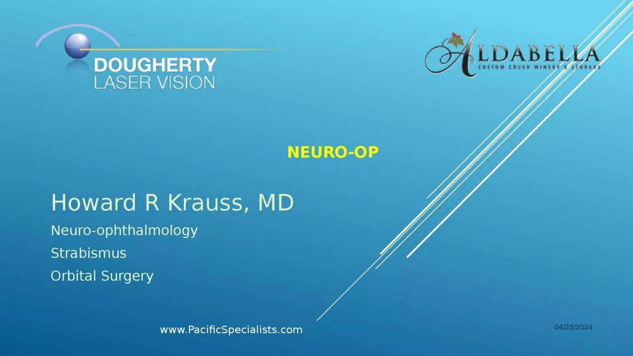
Neuroophthalmology Strabismus Orbital Surgery 412015 wwwPacificSpecialistscom nothing to disclothes Howard R Krauss MD Los Angeles CA neuroop Howard R Krauss MD ID: 918004

www pacificspecialists 2015 reduced pacificspecialists www reduced 2015 assess visual reassess approaches diagnostic patient optic neuro acuity field visionif

Share:

Link:

Embed:

Download Presentation from below link

Download Presentation The PPT/PDF document "neuro-op Howard R Krauss, MD" is the property of its rightful owner. Permission is granted to download and print the materials on this web site for personal, non-commercial use only, and to display it on your personal computer provided you do not modify the materials and that you retain all copyright notices contained in the materials. By downloading content from our website, you accept the terms of this agreement.


Presentation Transcript

Slide1

neuro-op

Howard R Krauss, MDNeuro-ophthalmologyStrabismusOrbital Surgery

4/1/2015

www.PacificSpecialists.com

Slide2

… nothing to disclothes …

Howard R Krauss, MDLos Angeles, CA

Slide3

neuro-op

Howard R Krauss, MDNeuro-ophthalmologyStrabismusOrbital Surgery

4/1/2015

www.PacificSpecialists.com

Pacific Eye & Ear

11645 Wilshire Blvd., Suite 600

Los Angeles, Ca. 90025

310-477-5558

DrKrauss@PacificSpecialists.com

www.PacificSpecialists.com

Slide4

pacific eye & ear

4/1/2015 www.PacificSpecialists.com

Pacific Eye & Ear is an association of eleven doctors, providing medical and surgical services encompassing Ophthalmology, ENT, Facial Plastic Surgery and Audiology.

Slide5

Diagnostic approaches to reduced vision1) Talk with and examine the patient

4/1/2015www.PacificSpecialists.com

Slide6

Diagnostic approaches to reduced visionWhen the vision is subnormal, proceed to:

2) Pinhole acuity 3) Refraction 4) Visual field assessment4/1/2015www.PacificSpecialists.com

Slide7

Diagnostic approaches to reduced visionIf corrected acuity is normal

and visual field is normal: 1) Complete the general examination and if all else is normal, proceed to discussion of optical services, from spectacles to contact lenses to surgery.4/1/2015www.PacificSpecialists.com

Slide8

Diagnostic approaches to reduced visionIf corrected acuity is abnormal or visual field is abnormal:

1) Proceed with Retinal Evaluation and/or consultation.4/1/2015www.PacificSpecialists.com

Slide9

Diagnostic approaches to reduced visionIf Retinal Consultant detects abnormalities and arranges treatment for same:

1) Re-evaluate patient to assess whether or not the retinal abnormalities are likely the only source of the patient’s complaints.4/1/2015www.PacificSpecialists.com

Slide10

Diagnostic approaches to reduced visionIf Retinal Consultant finds the retina to be normal, re-evaluate patient:

1) Reassess the tear film, cornea, crystalline lens, lens implant and posterior capsule as potential sources of reduced acuity; 2) Reassess the visual field reliability and pattern of abnormality 3) Assess relative light and color brightness and check for RAPD 4) Assess the optic nervehead appearance and (a)symmetry 5) Consider RNFL and RGC layer thickness analyses

6) Consider ERG 7

) Consider Neuro-ophthalmologic consultation.4/1/2015

www.PacificSpecialists.com

Slide11

Diagnostic approaches to reduced visionIf Retinal Consultant finds the retina to be normal, re-evaluate patient:

1) Reassess the tear film, cornea, lens implant and posterior capsule as potential sources of reduced acuity; 2) Reassess the visual field reliability and pattern of abnormality 3) Assess relative light and color brightness and check for RAPD 4) Assess the optic nervehead appearance and (a)symmetry 5) Consider RNFL and RGC layer thickness analyses

6) Consider ERG 7) Consider Neuro-ophthalmologic consultation.

4/1/2015

www.PacificSpecialists.com

Slide12

Diagnostic approaches to reduced visionIf Retinal Consultant finds the retina to be normal, re-evaluate patient:

1) Reassess the tear film, cornea, lens implant and posterior capsule as potential sources of reduced acuity; 2) Reassess the visual field reliability and pattern of abnormality 3) Assess relative light and color brightness and check for RAPD 4) Assess the optic nervehead appearance and (a)symmetry 5) Consider RNFL and RGC layer thickness analyses

6) Consider ERG 7) Consider Neuro-ophthalmologic consultation.

4/1/2015

www.PacificSpecialists.com

Slide13

Diagnostic approaches to reduced visionIf Retinal Consultant finds the retina to be normal, re-evaluate patient:

1) Reassess the tear film, cornea, lens implant and posterior capsule as potential sources of reduced acuity; 2) Reassess the visual field reliability and pattern of abnormality 3) Assess relative light and color brightness and check for RAPD 4) Assess the optic nervehead appearance and (a)symmetry 5) Consider RNFL and RGC layer thickness analyses

6) Consider ERG 7) Consider Neuro-ophthalmologic consultation.

4/1/2015

www.PacificSpecialists.com

Slide14

Diagnostic approaches to reduced visionIf Retinal Consultant finds the retina to be normal, re-evaluate patient:

1) Reassess the tear film, cornea, lens implant and posterior capsule as potential sources of reduced acuity; 2) Reassess the visual field reliability and pattern of abnormality 3) Assess relative light and color brightness and check for RAPD 4) Assess the optic nervehead appearance and (a)symmetry 5) Consider RNFL and RGC layer thickness analyses

6) Consider ERG 7) Consider Neuro-ophthalmologic consultation.

4/1/2015

www.PacificSpecialists.com

Slide15

Diagnostic approaches to reduced visionIf Retinal Consultant finds the retina to be normal, re-evaluate patient:

1) Reassess the tear film, cornea, lens implant and posterior capsule as potential sources of reduced acuity; 2) Reassess the visual field reliability and pattern of abnormality 3) Assess relative light and color brightness and check for RAPD 4) Assess the optic nervehead appearance and (a)symmetry 5) Consider RNFL and RGC layer thickness analyses

6) Consider ERG 7) Consider Neuro-ophthalmologic consultation.

4/1/2015

www.PacificSpecialists.com

Slide16

Diagnostic approaches to reduced visionIf Retinal Consultant finds the retina to be normal, re-evaluate patient:

1) Reassess the tear film, cornea, lens implant and posterior capsule as potential sources of reduced acuity; 2) Reassess the visual field reliability and pattern of abnormality 3) Assess relative light and color brightness and check for RAPD 4) Assess the optic nervehead appearance and (a)symmetry 5) Consider RNFL and RGC layer thickness analyses

6) Consider ERG 7

) Consider Neuro-ophthalmologic consultation.

4/1/2015www.PacificSpecialists.com

Slide17

Ocular Coherence Tomography (OCT)Neuro-ophthalmic Applications

Evaluation and Monitoring: MS / Optic Neuritis Ischemic Optic Neuropathy Any Optic Neuropathy Compressive Optic Neuropathy Papilledema

Slide18

55-year-old womanwith MSBCVA 20/30 OD 20/25 OS

Slide19

47-year-old Hawaiian womanAware of diminishing vision of the left eye over 1 year, rapidly worsening over the last 3 months.Intermittent mild pain OS, especially when flying.

www.PacificSpecialists.com

Slide20

Visual Acuity 20/25 OD 20/50-1 OS

No proptosisNo enophthalmosNo hyper- or hypoglobusOrthophoric in all positionsFull ductions2+ RAPD OS

www.PacificSpecialists.com

Slide21

Humphrey 10-26-11

www.PacificSpecialists.com

Slide22

Octopus 12-27-11

www.PacificSpecialists.com

Slide23

RNFL thkns 106 OD, 93 OS

www.PacificSpecialists.com

Slide24

www.PacificSpecialists.com

Slide25

Transnasal Image-Guided Orbital Surgery (TIGOS) TIGOS has been carried out by

Drs. Krauss & Griffiths since 2001. The work was presented at the 5th International Congress of the World Federation of Skull Base Societies in 2008.

4/1/2015

www.PacificSpecialists.com

Slide26

Outpatient Surgery

www.PacificSpecialists.com

Slide27

Image-guided Endoscopic Sx

www.PacificSpecialists.com

Slide28

www.PacificSpecialists.com

Slide29

www.PacificSpecialists.com

Slide30

Pre-op / Octopus / Post-op

www.PacificSpecialists.com

Slide31

Post-op

www.PacificSpecialists.com

Slide32

2 weeks post-op

UCVA 20/25Trace RAPD OSMild weakness of left adduction and infraduction – improving day-by-day

www.PacificSpecialists.com

Slide33

www.PacificSpecialists.com

Slide34

www.PacificSpecialists.com

Slide35

mri of the visual afferent systemBrain and Orbits with and without contrast

4/1/2015www.PacificSpecialists.com

Slide36

mri of the visual afferent systemIf you know the lesion is

retrogeniculate:Brain with and without contrast4/1/2015www.PacificSpecialists.com

Slide37

mri of the visual afferent systemIf you know the lesion is anterior visual pathway:

Orbits and pituitary with and without contrast4/1/2015www.PacificSpecialists.com

Slide38

BSB 54yo female

11/05: Puffiness OS

Va 20/15,20/25

Ext: H 16/21

P: 1.2log LAPD

EOM: min

↓ L elev

Slide39

BSB – W/U

OCT NFL (11/05):

Slide40

BSB – W/U

MRI (12/05):

Slide41

BSB – F/U

MRI (5/06):

Slide42

BSB – F/U

10/06: Diplopia in

right gaze

Va 20/20 OU

Ext: H 16/14

EOM: min

↓ L add

P: .3log LAPD

Slide43

BSB – W/U

OCT NFL (10/06):

Slide44

JWD 63yo male

3/06:

↓Va OS

Va 20/20,20/60

P: .9log LAPD

Slide45

JWD – POH

12/05: Routine check vision

Dx: “cataracts”

Referred for cataract extraction

Ophthalmologist said “no cataract”

Slide46

JWD – W/U

OCT:

Slide47

JWD – F/U

8/07: “No

Δ

Va 20/25 OU

P: .9log LAPD

Slide48

JWD – W/U

OCT NFL (8/07):

Slide49

KH 48yo female

11/08:

↓Va

Va 20/30,8/200

VF:

Ext: w/q

P: .3log LAPD

EOM: full

SLE: wnl

Fundus: nl DMV

4/1/2015

www.PacificSpecialists.com

Slide50

KH – PMH

1/08: Polydipsia

4/08: Amenorrhea

10/08: HA, N/V

4/1/2015

www.PacificSpecialists.com

Slide51

KH – W/U

OCT NFL (11/08):

4/1/2015

www.PacificSpecialists.com

Slide52

KH – W/U

MRI (11/08):

4/1/2015

www.PacificSpecialists.com

Slide53

KH – Rx

11/08:

Transphenoidal

endoscopic decompression

Path:

craniopharyngioma

4/1/2015

www.PacificSpecialists.com

Slide54

KH – F/U

8/09: “Better”

Va 20/20 OU

N 3pt OU

VF:

Ext: w/q

P: w/o APD

EOM: full

SLE: wnl

Ta: 19/22

Fundus:

4/1/2015

www.PacificSpecialists.com

Slide55

KH – W/U

OCT NFL (8/09):

4/1/2015

www.PacificSpecialists.com

Slide56

In summary:Listen

to the patient and solicit information.Examine the patient: determine BCVA and assess VF.Understand and explain symptoms and findings.Consider and recommend additional testing, or consultation, as indicated.Follow-up on all tests and consultations with patient.

Avoid contributing to a delay in diagnosis and treatment.

Slide57

neuro-op

Howard R Krauss, MDNeuro-ophthalmologyStrabismusOrbital Surgery

4/1/2015

www.PacificSpecialists.com

Pacific Eye & Ear

11645 Wilshire Blvd., Suite 600

Los Angeles, Ca. 90025

310-477-5558

DrKrauss@PacificSpecialists.com

www.PacificSpecialists.com