/
studying the optics of the lens becausein most casesthe lens is studying the optics of the lens becausein most casesthe lens is

studying the optics of the lens becausein most casesthe lens is - PDF document

davies
davies . @davies
Follow
343 views
Uploaded On 2022-09-01

studying the optics of the lens becausein most casesthe lens is - PPT Presentation

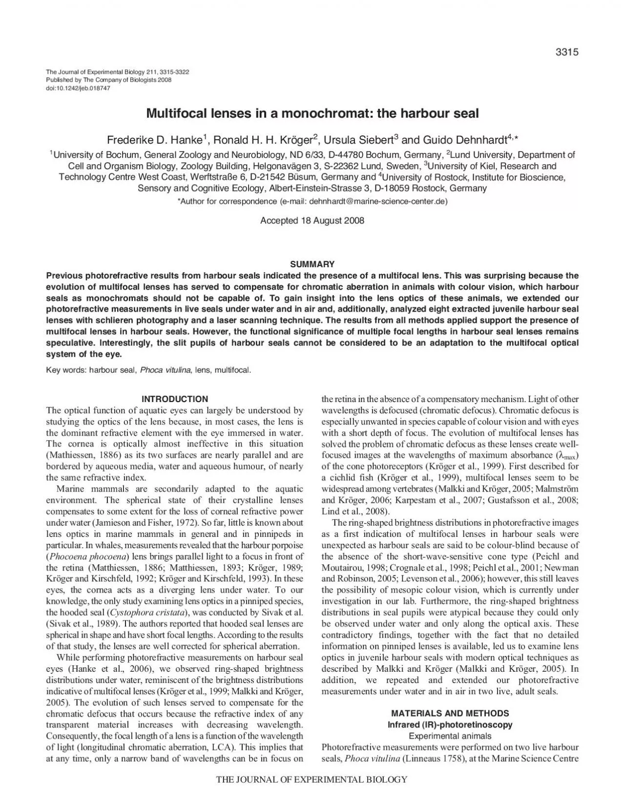
3315 The Journal of Experimental Biology 211 33153322Published by The Company of Biologists 2008Multifocal lenses in a monochromat the harbour sealFrederike D Hanke1 Ronald H H Kröger2 Ursula ID: 945828

seal fig lenses lens fig seal lens lenses harbour 133 seals rings centre approximately distance laser eye schlieren water

Share:

Link:

Embed:

Download Presentation from below link

Download Pdf The PPT/PDF document "studying the optics of the lens becausei..." is the property of its rightful owner. Permission is granted to download and print the materials on this web site for personal, non-commercial use only, and to display it on your personal computer provided you do not modify the materials and that you retain all copyright notices contained in the materials. By downloading content from our website, you accept the terms of this agreement.


Presentation Transcript

3315 studying the optics of the lens because,in most cases,the lens is The Journal of Experimental Biology 211, 3315-3322Published by The Company of Biologists 2008Multifocal lenses in a monochromat: the harbour sealFrederike D. Hanke1, Ronald H. H. Kröger2, Ursula Siebert3and Guido Dehnhardt4,*1University of Bochum, General Zoology and Neurobiology, ND 6/33, D-44780 Bochum, Germany, 2 3316 3years old; Sam, 12years old) were experimentally experiencedet al., 2006). The IR-photoretinoscope (Fig.1A, inset) consisted of50mm, †/1:1.4, Hamburg, Germany; with twoextension rings, resulting in an operating distance of 0.5m)of anIR-sensitive monochrome CCD camera(The Imaging Source,and retinoscope were placed at a distance of 0.5m in front of thein the lens aperture) (Fig.1A, inset) orientated horizontally. For(Fig.2). After lowering the head, the eyes were close to theapproximately two weeks (Table1). In Germany, harbour seals areare given in Table1 for all seals.lens and its close adherence to the vitreous (Fig.3B), the eye was(approximately5mm5mm) (Fig.3A) were left attached. Thisserved as a handle,which allowed the lens to be placed in the setups (Fig.3B), as is encountered in the young human eye (Sachse

nweger,The lens of the first eye was extracted between 0.5 and 2.5h afterthe death of the animal (Table1). The second eye was excised F. D. Hanke and others Immersion bathLight sourceCameraFocusingCameraImmersion bathLens holder AC RCR KE Fig.1. Schematic representation of the experimental setups used tomeasure harbour seal lenses. (A)IR-Photoretinoscopy under water. A(B)Schlieren photography. White light (point source, cold light laboratorylamp, 3200K) was reflected by a beam splitter into the optical axis. The7.4, 290mosmol, 20°C), focused the lightto which a small amount of microparticles was added. A 5mW green(537nm) diode-pumped, solid-state laser was used to scan through a 3317 Multifocal lenses in harbour seals running at 3200K) was reflected by a beam splitter into the opticalaxis (Fig.1B). The seal lens was suspended in an immersion bath7.4, 290mosmol,recording the immersion bath at a distance of 10cm from above(Fig.1C). The images long axis was slightly turned (approximately10deg.) relative to the laser beam in order to minimize aliasing7.4, 290mosmol, used for measuring fish lenses (Malkki and Kröger, 2005). A 5mWgreen (537nm) diode-pumped,solid-state laser was used to scan100mm lens withlaser

beam,which was scattered upwards by the microparticles.1ml of aqueous was extracted with a syringe. The syringe and its Fig.2. Harbour seal Enzo during underwater photorefractive Table 1. Age and life history of all animals used for schlieren photography and laser scanning, as well as time between death and start of the experiments in hoursTime between death and 2hours14days15days 17days 3318 estimated as 0.1mm. For the determination of refractive index andobtained under water (Fig.4A…C). However, extendedwhen the pupil is fully dilated (Fig.4A…C). Two faint rings arepresent at approximately 0.5lens radius (R) and 0.75R (Fig.4A).All rings are more pronounced in the adult seal (Fig.4C). In air,but can still be detected between approximately 0.65 and 0.75R(Fig.4D).(Fig.5). A red outermost ring and two bluish rings are clearly present ring was thin and incomplete (Fig.5C). The overall pattern is variableinhomogenously coloured (e.g. Fig.5C). Especially in the centre,two seals (Fig.5A,B), there are soft transitions between the rings.Pictures of the lens of seal 1 (Fig.5A) indicate that the lens is slightlylenses of seal 4 (Fig.5D), the respective schlieren photographs arewater (Fig.4A) with the two blui

sh rings correlating to the two moreprominent ring at approximately 0.85R. The lens sutures areprominent on all schlieren photographs (Fig.5). They stretch overalmost 0.8R and are of cross-like appearance.The results are irregular for the central BCDs between 0 and 0.3R(Fig.6A…D) where the accuracy of the method is low (Malkki andof the newborn (Fig.6A), show two peaks in the periphery(Fig.6B…D, long arrows). Furthermore, a sharp decline in BCD(Fig.6A…D, short arrows). The mean LSA curve of all, except forthe lenses of the neonate, is presented in Fig.7A. Two peaks ofslightly different BCDs can be seen (first peak at BEP 0.67R, BCD3.77R; second peak at BEP 0.87R, BCD 3.75R). The mean BCDbetween 0.3 and 0.6R is 3.58R. BCD increases minimally towardsthe centre by 0.05R (1.4% of mean value 3.58R). At 0.9R, BCD F. D. Hanke and others Fig.3. Dissection of harbour seal lenses. (A)Lens lyingiris, and after cutting the zonular fibre ring. (B)Posteriorattached zonular fibres and the lens. Scale bar, 5mm. BCD Fig.4. Photorefractive images of a young (3years old) harbour seal under water axially refracted with lateral (A) and frontal camera position (B) and an older(12years old) harbour seal under water (C) and i

n air (D). On the underwater pictures (A…C), two rings in the centre and one pronounced ring in theperiphery can be seen. Corneal cloudiness in the older seal leads to a central stripe in the brightness distribution under water (C), and irregularities in 3319 Multifocal lenses in harbour seals drops steeply by approximately0.5R (14% of mean value 3.58R)before increasing again up to values of approximately 5R. The meanLSA curve (Fig.7A) mirrors the results of photorefractivemeasurements in the live seals under water (Fig.4A…C) and theschlieren photographs of the best dissected lens (Fig.5D) as againtwo broad rings and a thin sharp ring can be discerned clearly. Fig.7Bosmolarity measurements are listed in Table2. Due to the dissection33.0±0.08mm(Table2). The equatorial diameter of the eye-cup wasslightly larger in older animals (Table2).Mean axial lens diameter (±s.d.)is 10.90±0.19mm, mean equatorialdiameter (±s.d.)11.69±0.33mm (Table2). 6; Table2). Mean osmolarityof both media is 343.6mosmol (aqueous, 344.571±22.397mosmol,7; vitreous, 342.667±12.127mosmol, 6; Table2).irregularities have an effect on the brightness profiles in air (Fig.4D) Fig.5. Results of schlieren photography. For each seal, one typ

ical picture is presented in chronological order of measurement (A,picture of seal 1; B, seal 00.20.40.60.81Beam entrance position (R)Back centre distance (R) 00.20.40.60.81 3.43.53.63.73.83.94.03.63.73.84.04.14.23.43.53.63.73.83.94.03.43.53.63.83.94.0 Fig.6. Results of laser scanning displaying the longitudinal spherical aberration (LSA) curves of harbour seal lenses measured at 537nm. (A…D) LSA curvesof the right eyes of seal 1 (A), seal 2 (B), seal 3 (C) and seal 4 (D). Long arrows mark peaks, short arrows mark steep declines in back centre distance. 3320 navigate independently.The juvenile lenses we studied werethat the osmolarity in seal eyes is approximately 53mosmol higher focal length than other regions of the lens (Fig.6A), which suggeststhe laser scanning method for BEPs smaller than 0.3R (Malkki andThe LSA curve obtained by averaging all scanning results (Fig.7A) F. D. Hanke and others 00.20.40.60.81 Beam entrance position (R)Back centre distance (R) Fig.7. (A)Mean LSA curve of all lenses except for the neonates lenses. Two peaks at 0.67R and 0.87R are visible (long arrows). Note the steep decline inback centre distance in the periphery (short arrow). (B)An example picture of a spherical harbour s

eal lens deflecting the laser beams. Scale bar, 5mm. Table 2. Eye and lens dimensions, refractive index and osmolarity of the aqueous and vitreous humours Eye diameter (mm)Lens diameter (mm)AqueousVitreous Refractive indexOsmolarity Refractive index Osmolarity SealEye EquatorialAxialEquatorial10.7412.0410.8911.5611.0811.3410.5411.2111.1311.5210.8911.8410.9511.9910.9612.0410.9011.69 ±0.19±0.33±0.00010±12.127 3321 Multifocal lenses in harbour seals seals (Fig.4). Furthermore, there are two peaks in the mean LSAcurves at approximately 0.67R and 0.87R (Fig.7A). These peakscorrespond well to bluish rings on schlieren photographs (Fig.5), theouter one of these being narrow in the lenses of seal 3 (Fig.5C). Intwo if light intensity increases from 0.1cdmalmost circular) to 80cdmposition of 0.67R and 0.87R and both contribute to the image if thepupil is fully dilated (radius 1R). However, at 80cdm Phoca largha(Wartzok and McCormick, 1978); two species of furseals, Arctocephalus pusillusand Arctocephalus australis(Busch andDücker, 1987); California sea lion, Zalophus californianus(Griebeland Schmid, 1992)] have revealed some colour vision in theblue…green range of the spectrum. However, except for the study oncolour

vision in the Bering sea spotted seal (Wartzok and McCormick,1978), the seals might have used brightness instead of colour cuesifthe seals sensitivity for brightness differences had beenlies at 495…501nm (Lavigne andis assumed to be approximately 550nm, assessed as 510nm, might be explained by an overlap of theThis overlap would have shifted the spectral sensitivity peak to 510nmto be 496nm (Lavigne and Ronald, 1975) and 550nm, respectively,nm, respectively,öCampbell (Kröger and Campbell, 1996)], which would makemultifocal optical systems beneficial. Harbour seal lenses thereforesolve the problem of chromatic aberration in dim light. In bright light,where the pupil is almost completely or fully constricted, depth offocus is increased and there is little chromatic blur. The difference infocal length would be 0.6% in the case of the rod and cone maxat496nm and 510nm (Crognale et al., 1998), respectively. Such a small 3322 BCDback centre distanceBEPbeam entrance positionIRinfraredLCAlongitudinal chromatic aberrationLEDlight emitting diodeLSAlongitudinal spherical aberrationPBSphosphate buffered salineRlens radiusTIFFtagged image file format (2003). Animal colour vision …behavioural F. D. Hanke and