/
xperience With Ultrasonography in the iagnosis and reatment xperience With Ultrasonography in the iagnosis and reatment

xperience With Ultrasonography in the iagnosis and reatment - PDF document

davis
davis . @davis
Follow
342 views
Uploaded On 2022-08-31

xperience With Ultrasonography in the iagnosis and reatment - PPT Presentation

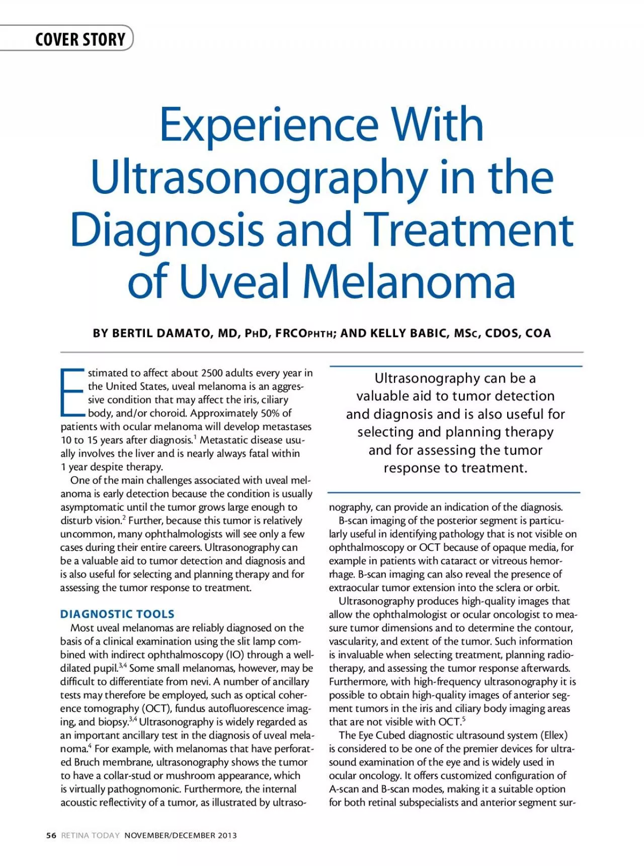
N 2013 BY BERTIL DAMATO MD PD FRCOHTH KELLY BA MS CDOS COA E Ultrasonography can be a valuable aid to tumor detection and diagnosis and is also useful for selecting and planning therapy and for ID: 944114

scan lesion figure eye lesion scan eye figure ultrasound tumor probe patient scans diagnosis internal nerve optic 146 ocular

Share:

Link:

Embed:

Download Presentation from below link

Download Pdf The PPT/PDF document "xperience With Ultrasonography in the ia..." is the property of its rightful owner. Permission is granted to download and print the materials on this web site for personal, non-commercial use only, and to display it on your personal computer provided you do not modify the materials and that you retain all copyright notices contained in the materials. By downloading content from our website, you accept the terms of this agreement.


Presentation Transcript

N 2013 xperience With Ultrasonography in the iagnosis and reatment BY BERTIL DAMATO, MD, PD, FRCOHTH KELLY BA MS, CDOS, COA E Ultrasonography can be a valuable aid to tumor detection and diagnosis and is also useful for selecting and planning therapy and for assessing the tumor response to treatment. COVER STORY OVEMBE 2013RETINA geons. Cataract surgeons benefit from the inclusion of an immersion A-scan feature, which eliminates corneal compression and transmits ultrasound waves through dense cataract. Furthermore, the 40 MHz UBM widefield anterior segment B-scan mode gives ophthalmologists an unprecedented view of the lens apparatus, including zonules and ciliary muscles (areas that are almost impossible to visualize with other ultrasound systems). The 10 MHz posterior B-scan mode allows visualization of deeper tissue structures in the posterior segment. Such level of detail is possible thanks to the Eye Cubed’s signal-to-noise ratio, which minimizes noise and allows the system to detect echoes from some of the smallest ocular structures. The Eye Cubed also features real-time high-resolution imaging and advanced movie mode using the fastest sampling rate available. It also has an internal memory for storing scans and is DICOM compatible, so data can be easily cross-referenced with patient details and transferred to an electronic medical record to optimize practice management. Figure 1. Precise ultrasonographic measurements of tumor dimensions and distance from optic nerve enabled accurate proton beam radiotherapy, successfully conserving the eye with good vision.Figure 2. Dome-shaped melanoma superior to the right optic disc (A). Longitudinal and transverse B-scans provide accurate measurements (B and C). A-scan ultrasonography demonstrates typical internal acoustic reflectivity (D). N 2013 One of us (BD) has used the Eye Cubed for more than 20 years, first at the Liverpool Ocular Oncology Centre in England and, more recently, at the Ocular Oncology Service of the University of California, San Francisco. This equipment has proved invaluable for diagnosis, treatment planning, and tumor monitoring. To follow are some cases in which the Eye Cubed proved particularly helpful.Using the Eye Cubed, we were able to identify a very rare extraocular local recurrence adjacent to the optic nerve following plaque radiotherapy of a medial choroidal melanoma in the right eye. Precise ultrasonographic measurements of tumor dimen

sions and distance from optic nerve enabled accurate proton beam radiotherapy, successfully conserving the eye with good vision (Figure 1). Figure 2A shows a dome-shaped melanoma superior to the right optic disc. Longitudinal and transverse B-scans provide accurate measurements (Figures 2B and 2C). A-scan ultrasonography demonstrates typical internal acoustic reflectivity (Figure 2D).Figure 3 shows a small peripheral cilary body mass lesion. The dimensions and extent of the lesion were well demonstrated with the Eye Cubed’s B-scan probe.Figure 4A shows a superotemporal choroidal hemangioma in the right eye of a 9-year-old boy. EyeCubed A- and B-scans show high internal reflectivity consistent with this diagnosis (Figure 4B and 4C). The Eye Cubed is a whole-eye solution to ultrasound imaging, with a wide range of scans and adjustments. It is, however, essential for the examiner to have proper training on how to use the technology correctly. Without adequate expertise, it is difficult to get the best out of any machine, but this is particularly true with ophthalmic ultrasound. There is also a danger of misinterpreting the information provided, resulting in an incorrect diagnosis. When planning treatment or assessing tumor growth or regression over time, it is essential to measure tumor dimensions accurately and consistently. For example, when measuring tumor thickness, it is important to identify the interface between tumor and sclera and essential to measure the thickest part of the tumor with the probe held perpendicular to the internal scleral surface. Without good technique, the examiner may conclude that the tumor has grown and send the patient for emergency treatment, thus causing the patient unnecessary distress and perhaps Figure 3. Ciliary body tumor with cystic spaces clearly defined by ultrasonography. Figure 4. Nine-year-old boy with a superotemporal choroidal hemangioma in the right eye (A). EyeCubed A- and B-scans show high internal reflectivity consistent with this diagnosis (B and C). OVEMBE 2013RETINA By Rhonda G. Waldron, MMSc, COMT, CRA, ROUB, Ultrasound imaging is essential in ocular oncology, guiding both the diagnosis and management of intraocular tumors. The following is designed as a primer to provide important tips for obtaining the best possible ultrasound images. Examine the patient with the eye . The lid absorbs ultrasound energy, resulting in loss of information. Additionally, both the prob

e face and marker orientations must be exact and are dependent on patient gaze. The eyes roll up when the lids are closed, making it extremely hard for the patient to look in appropriate gazes (particularly down gaze) as needed for the exam. Proper localization of the lesion will be much faster when gaze is certain, and better resolution of the images will be obtained when the probe is placed on 0.3% gel duct the sound into the eye. Regular ultrasound gels are corneal irritants and should never be used in ophthalmology. Methylcellulose can be used, but it isn’t as thick as HPMC, it must be rinsed out afterward, and a significant percentage of the patient population will be sensitive to its preservative. When scanning a lesion, superimpose a clock around the eye being examined. Direct the patient’s gaze toward the area of interest, with the probe face on the conjunctiva opposite the area of interest to center the pathology in the echogram where best resolution is obtained. Perpendicularity to the vertex of the lesion is achieved when the vertex appears brighter than the Perform 1 transverse (lateral) scan of the lesion on high gain to document any vitreal debris (Figure 1)verse scans, the marker should be parallel to the limbus and nasal in orientation if scanning either 12 o’clock or 6 o’clock, and superior in orientation for any other clock Turn the gain down for better resolution of the lesion and perform 4 more transverse scans of the lesion, sweeping the scan line back and forth across the lesion est elevation. Compare the height and basal dimensions between the 4 scans, erring toward the largest measurements to ensure adequate treatment. IMPROV Figure 1. Transverse scan of a choroidal melanoma centered at 5:00, OD. Note the brightening of the lesion’s vertex, indicating perpendicularity as well as the associated subretinal fluid extending from the edges of the lesion.Figure 3. Axial scan of same lesion. Note that the posterior lens surface is centered to the left of the echogram, and the optic nerve shadow is centered to the right of the echogram. Because the lesion is located inferiorly at 5:00, the probe marker was oriented at 11:00 so that the lesion appears below the optic nerve.Figure 4. Diagnostic A-scan of the same lesion. Note the retinal and scleral echoes are tall, with the spikes representing the internal reflectivity of the lesion being low-to-medium in height.Figure 2. Longitudinal

scan of same lesion centered at 5:00. To view the anterior border of this large lesion, the probe was shifted toward the fornix, limiting the visibility of the optic nerve shadow below. N 2013 inappropriate therapy. Further, although many ophthalmologists have great faith in ultrasonography as a method of diagnosing ocular tumors, the use of ancillary tools to support or confirm a diagnosis should not be ruled out. For example, if a particular diagnosis is unclear, or if a patient presents with a rare condition, a biopsy may be necessary to ensure a correct diagnosis. Ultrasound imaging is an important tool in ocular oncology and is increasingly regarded as the most important ancillary test, after slit-lamp examination and ophthalmoscopy, in the diagnosis of ocular melanoma. This is because ultrasound allows eye specialists to view structures that cannot otherwise be assessed. Ultrasonography also produces high-quality images that allow the ophthalmologist or ocular oncologist to measure the size, shape, and extent of tumors. Ultrasound is a quick and convenient means of obtaining excellent images of the posterior segment when there is a cloudy cornea, dense cataract, or vitreous hemorrhage. Adequate training, however, is essential to reap the benefits ultrasound can offer in the diagnosis and treatment of uveal Bertil Damato, MD, PhD, FRCOphth, is Director of Ocular Oncology Service and Professor of Ophthalmology and Radiation Oncology at the University of California, San Francisco, Medical Center. He may be reached at +1 415 514-6918; or at DamatoB@Vision.UCSF.edu. Kelly Babic, MSc, CDOS, COA, is an Ophthalmic Ultrasonographer and the Diagnostic Services/Clinic Manager at University of California, San Francisco, Medical Center. She may be reached at Kelly.Babic@UCSFmedctr.org. Perform 4 longitudinal (radial) scans of the lesion on lowered gain with the probe marker now directed perpendicular to the limbus, directly at the clock hour being examined (Figure 2). Make certain the optic nerve shadow is visible in the lower right portion of the echogram, with the lesion centered in the area of best resolution. Note: If a lesion extends to the far periphery this centration may not be possible, and the optic nerve shadow may be so low on the right hand side that it can’t be displayed on the echogram. It is also important in lesions located or extending into the far periphery that ultrasound biomicroscopy be performed to rul

e out ciliary body involvement. Measure the height and basal dimensions again among the 4 scans. The height should correspond to the height eter once again to ensure adequate treatment. Perform an axial scan (if the eye is not pseudophakic) by changing the patient’s gaze to primary and centering the probe face on the corneal vertex (Figure 3). Correct alignment is indicated when the posterior lens surface is centered toward the left of the echogram gram. The marker orientation is determined by the location of the lesion. For lesions located superiorly or nasally, aim the marker directly toward the lesion and it will appear above the nerve in the image. For lesions located temporally or inferiorly, aim the marker opposite the lesion and it will appear below the nerve in the image. Document the size of the lesion to the nearest 0.1 mm by writing the greatest height first, followed by the greatest transverse base, and finally A diagnostic A-scan should also be performed to ascertain the internal reflectivity of the lesion (Figure 4). This is done with a calibrated, dedicated A-scan probe available from a few ultrasound manufacturers. (The cross vector placed on top of the B-scan image is NOT a calibrated A-scan and should not be used for diagnostic purposes.) The patient’s gaze is still directed toward the area of interest, with the diagnostic A-scan probe placed on the opposite conjunctiva. Correct alignment is assured when the retinal spike is tall and straight, as is the spike representing the sclera. When using the gain setting as per the pared to known lesion patterns for diagnostic purposes. Melanomas tend to have low-to-medium, regular internal reflectivity, metastatic carcinomas tend to have medium-to-high, irregular internal reflectivity, and hemangiomas tend to have high, regular internal reflectivity.Vascularity may be more difficult to see on diagnostic A-scan exams with digital equipment, but many times it is easy to see on the B-scan exam. Center the lesion in the echogram with gain lowered and hold the probe steady, observing the center of the lesion. Vascularity is appreciated as areas of pulsation within the lesion. When visualized, Rhonda Waldron is a Diagnostic Echographer and Senior Associate in Ophthalmology at Emory Eye Center at Emory University in Atlanta. She also owns Eye Scan Consulting, and teaches both A-scan and B-scan ultrasound. She can be reached at rwaldro@emory.edu. COVER STORY COVER S