PPT-GRANULOME PÉRIAPICAL
Author : debby-jeon | Published Date : 2016-08-08
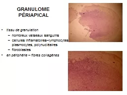
tissu de granulation nombreux vaiseaux sanguins cellules inflamatoireslymphocytes plasmocytes polynucléaires fibroblastes en périphérie fibres collagènes KYSTE
Presentation Embed Code
Download Presentation
Download Presentation The PPT/PDF document "GRANULOME PÉRIAPICAL" is the property of its rightful owner. Permission is granted to download and print the materials on this website for personal, non-commercial use only, and to display it on your personal computer provided you do not modify the materials and that you retain all copyright notices contained in the materials. By downloading content from our website, you accept the terms of this agreement.
GRANULOME PÉRIAPICAL: Transcript
Download Rules Of Document
"GRANULOME PÉRIAPICAL"The content belongs to its owner. You may download and print it for personal use, without modification, and keep all copyright notices. By downloading, you agree to these terms.
Related Documents