/
31212019182417161515241420rf 31212019182417161515241420rf

31212019182417161515241420rf - PDF document

deena
deena . @deena
Follow
342 views
Uploaded On 2022-08-25

31212019182417161515241420rf - PPT Presentation

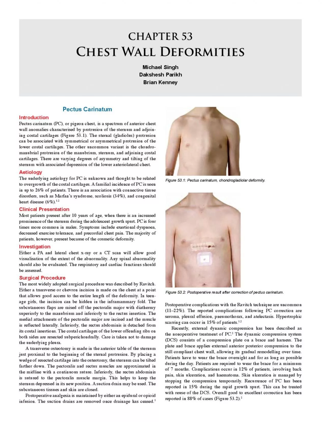
Figure 531 Pectus carinatum chondrogladiolar deformity 30292827263125242323312228212019181726172827 Figure 533 Pectus excavatum ID: 941502

bar chest figure sternal chest bar sternal figure sternum patients wall syndrome pectus defect excavatum procedure ribs deformity depression

Share:

Link:

Embed:

Download Presentation from below link

Download Pdf The PPT/PDF document "31212019182417161515241420rf" is the property of its rightful owner. Permission is granted to download and print the materials on this web site for personal, non-commercial use only, and to display it on your personal computer provided you do not modify the materials and that you retain all copyright notices contained in the materials. By downloading content from our website, you accept the terms of this agreement.


Presentation Transcript

\r\f \n\t\tPectus carinatum (PC), or pigeon chest, is a spectrum of anterior chest wall anomalies characterised by protrusion of the sternum and adjoining costal cartilages (Figure 53.1). The sternal (gladiolus) protrusion can be associated with symmetrical or asymmetrical protrusion of the lower costal cartilages. The other uncommon variant is the chondromanubrial protrusion of the manubrium, sternum, and adjoining costal Figure 53.1: Pectus carinatum, chondrogladiolar deformity.  Figure 53.3: Pectus excavatum severe deformity. Pectus excavatum (PE), or funnel chest, describes a posterior depression of the lower sternum and costal cartilages into the thoracic cavity. Cosmetic appearance is the main presenting reason in asymptomatic patients. The asymmetrical depression is not unusual, and is associated with sternal torsion.Pectus excavatum is an uncommon abnormality with an incidence of 38 per 10,000 and 7 per 10,000 births in the Caucasian and African populations, respectively. It is four times more common in males than females. A family history of PE is reported in 43% of patients, and familial association is seen in 7% of patients with pectus carinatum .The incidence of PC in our experience is equal to that of PE; however, the literature suggests that it is less common than PE.The sternal depression is thought to result from asymmetrical growth of the costochondral cartilages. However, the exact aetiology is unknown. There is an association with connective tissue disorders such as Marfan’s syndrome (21.5%) and Ehlers-Danlos syndrome (2%).Thirty percent of PE patients present in early childhood, with the majority presenting during the pubertal growth spurt. Common symptoms attributed to PE include exercise intolerance, dyspnoea, chest pain with and without exercise, and palpitations. The majority of patients are healthy, and they present because of the cosmetic appearance (Figure 53.3). Patients often have a slouched posture, and young children have an associated protuberant abdomen. Almost a quarter of PE cases are associated with scoliosis, and hence the spine should be investigated in all cases.Several variations in the sternal abnormality have been described. A cup-shaped appearance describes an abnormality with localised, steeply sloping walls. A saucer-shaped appearance is a diffuse and shallow sternal depression. A long asymmetrical trench-like deformity may also be found. Varying degrees of asymmetry of the chest wall may be present. Sternal torsion may be clinically obvious. A mixed carinatum/excavatum is an uncommon variation with the presence of a carinatum (protuberance) of the manubrium and excavatum of the sternum (Figure 53.4).Cardiac abnormalities have been known to be associated with PE and should be investigated in all cases with electrocardiography and echocardiography. Compression of the right atrium and ventricle by the depressed sternum has been implicated to cause mitral or tricuspid valve prolapse in up to 17% of patients. This has not been our experience in the United Kingdom; however, we have rarely found associated cardiac abnormalities in PE patients. Conduction abnormalities on electrocardiogram (ECG), such as right heart block, first-degree heart block, and Wolff-Parkins

on-White syndrome, may be present in up to 16% of patients.Respiratory function should be assessed preoperatively at least with spirometry. Restrictive lung functions have been reported, with decreases in forced vital capacity (FVC) of 77%, forced expiratory volume in 1 second (FEV) of 83%, and forced expiratory flow during the middle portion of expiration (FEFof 73%. It is useful to identify any underlying respiratory abnormalities prior to surgery, both for anaesthesia and to see whether correction results in improvement. Chest x-rays, both anteroposterior (AP) and lateral, are routinely performed, which may help to define the severity of the deformity as well Figure 53.5: Chest x-ray AP view showing shift of heart towards left and a line as help to evaluate the thoracic spine (Figures 53.5 and 53.6). A computed tomography (CT) scan of the chest has been considered the gold standard investigation in PE (Figure 53.7). It allows the calculation of the Haller index, the ratio of the transverse to the AP diameter at the lowest point of the depression. Other information obtained includes the length of the depression, degree of sternal torsion, and presence of chest wall asymmetry. Although recommended by some, we do not routinely carry out a CT scan of the chest or calculate the Haller index, as they do not influence the operative technique or outcome.Surgery for PE is carried out mainly to improve the appearance of the deformity. However, in some severe cases, two or more of the following are considered an indication for surgery:a Haller index of greater than 3.25 plus the presence of cardiac or pulmonary compression.Demonstrable cardiac abnormalities.Decreased pulmonary function; andPrevious failed repair by Ravitch or Nuss procedures.The Nuss procedure is ideally performed between 10 and 12 years of age, taking advantage of the pliability of the chest wall. The child in this age group, however, is often immature and unable to make an educated decision on whether to undergo such a major procedure mainly for cosmetic reasons. The patient should be Gillick competent to give informed consent. A single bar achieves good correction at this age. Two bars are recommended for the postpubertal patient, long or extensive depressions, or the presence of connective tissue disorders.The Ravitch procedure was the first widely accepted procedure for treatment of pectus excavatum. As has been described above for pectus carinatum, the cartilages are exposed and removed. The anterior table of the sternum is divided transversely to flatten the protrusion and the sternum is stabilized as described for the carinatum, but often with the addition of a transverse fixation bar, which is removed 6-12 months later. In the past decade, the minimally invasive repair (MIR), or Nuss procedure, as described by Dr. Donald Nuss, has become the most accepted technique for the correction of PE in developed countries.The patient is positioned supine with both arms abducted to 70° to 80° at the shoulder. Prophylactic intravenous antibiotics (e.g., cefuroxime) are given. An extensive skin preparation with an alcohol-based antiseptic solution of the anterior and lateral chest wall is essential.The distance is measured between the midaxillary points at the deepest part of the sternal depression. One inch is subtracted from

this measurement to determine the length of the bar. The bar is then bent symmetrically into a semicircular shape. It is important to have a 2–4 cm flat segment at the centre of the bar to support the sternum. A slight overcorrection is advisable.The most elevated point in line with the deepest point of excavatum on the costal ridges is marked (Figure 53.8). Transverse incisions are made across the midaxillary line at the level of the lowest point of the depression bilaterally. A subcutaneous tunnel is dissected to the top of the ridges. A 5-mm thoracoscope is inserted into the interspace inferior to the proposed site of bar insertion on the right side. A pneumothorax is maintained at a pressure of 5 to 7 mm Hg with a flow rate of 1–2 l/min. The rest of the procedure is performed under thoracoscopic visualisation.An introducer is then inserted from the midaxillary incision along the previously created subcutaneous tunnel, through the marked intercostal space (Figure 53.8). This introducer is used to carefully dissect the space between the sternum and pericardium under thoracoscopic vision. The introducer is brought out through the left symmetrically opposite, previously marked intercostal space. After passing the introducer through to the opposite midaxillary incision, both ends of the introducer are lifted measure for the Haller index by calculating AB/CD. while the costal margins and flared ribs are pushed down. This corrects the deformity and loosens up the connective tissue around the sternum. An umbilical tape is attached to the end of the introducer and pulled across the retrosternal space by withdrawing the introducer. The bent bar is attached to the end of the tape and pulled into the chest, across the mediastinum, and out through the left with the convexity facing posteriorly. The bar is then flipped by using bar flippers so that the convex surface is facing the sternum. This produces an instant correction of the depression.A single bar stabiliser is placed on the right side and sutured to the adjacent muscle with 1/0 polypropylene sutures. In addition, on the right side, the bar can be fixed to the adjacent rib with pericostal  Horner’s syndrome; transient, epidural relatedPericarditis (postcardiomyotomy syndrome); oral indomethacinPleural effusion; chest drainageTable 53.1: Early and late postoperative complications following bar insertion.1/0 polypropylene sutures guided by thoracoscopy. In the past, two stabilizers were used, one on either side. This practice has been abandoned by most because, as the chest wall grows, the patient can develop an hourglass deformity due to restriction in lateral growth, a problem that does not occur with the use of a stabilizer on only one side. The subcutaneous tissues and skin are closed. The lungs are expanded with positive pressures, and pneumothorax is relieved by putting a tube underwater through a thoracoscopy port site.A chest x-ray is obtained on day 1 postoperatively (Figure 53.9). Analgesia is maintained with epidural or a patient-controlled morphine infusion in combination with nonsteroidal anti-inflammatory drugs (NSAIDS). A graded programme of incentive spirometry and physiotherapy is commenced postoperatively. The epidural or morphine infusion is usually stopped after the third postoperative day. Oral NSAIDS and codeine

may be required for up to 3 weeks postoperatively. Patients are advised to avoid sporting activity for 3 months postoperatively. This allows sufficient scar tissue to develop around the bar, thus fixing it in place and preventing displacement.In experienced hands, surgical complications, summarised in Table 53.1, are uncommon. The majority of early postoperative complications can be managed conservatively. Late postoperative complications also are uncommon (see Table 53.1). Bar displacement is caused by inadequate fixation of the bar. Hence, it is recommended that the bar be fixed by using a bar stabiliser and pericostal sutures. Persistent postoperative pain should be investigated for bar or stabiliser displacement, a tight or too long bar, sternal or rib erosion, infection, and bar allergy (i.e., allergy to nickel).The bar is generally removed after 3 years. Under general anaesthesia, both lateral incisions containing the stabilizer bar is reopened. All visible sutures around the stabiliser are excised. The bar is straightened by using the bar reverse bender. Once reasonably loose, it is pulled out from the right side of the chest. The bar should not be forcibly extracted. In the event of difficulty, any residual scar tissue impinging on the bar should be excised before removal. Bar removal is generally uncomplicated. A postoperative chest x-ray should be obtained to rule out pneumothorax. Pneumothorax following bar removal usually is self-limiting and does not require any intervention.The long-term cosmetic results from the Nuss procedure are as follows: excellent, 86%; good, 10.3%; fair, 2.4%; and failed, 1.3% (see Figure 53.10).Poland’s SyndromePoland’s syndrome is a rare congenital malformation involving the chest wall and variable severity of other defects involving the areola, subcutaneous tissues, muscles, ribs, hand, and heart. The extent of these defects varies significantly from the absent sternocostal head of the pectoralis major and/or minor with normal breast and underlying ribs to complete absence of anterior portions of second to fifth ribs and cartilages. Breast involvement is frequent and is a disfiguring defect in girls. The hand deformity on the side of the defect is also associated in variable frequency from syndactyly to hypoplastic fingers.The reported incidence of Poland’s syndrome is low (1 in 30,000) and sporadic in nature. The exact aetiology of this defect is unknown. The proposed aetiology is a disruption in the subclavian arterial blood supply of the limb bud during the 6th foetal week.The anatomical abnormalities of Poland’s syndrome are usually unilateral. Clinically, these patients have an absent anterior axillary fold with the posterior axillary fold being easily visible from the front. The nipple and areola may be hypoplastic or absent with deficient subcutaneous tissues. The chest is depressed on the affected side due to hypoplasia or absence of the underlying 2–4 or 3–5 ribs and cartilages. Rarely, the lung may herniate through the defect in the chest wall, giving a flail segment. This may cause respiratory distress in the newborn period. Dextroposition of the heart is common in Poland’s syndrome, rather than dextrocardia. The surgical reconstruction options depend on the age of the patient and the extent of the defect. In the neonate, a flail chest may require reconstruction in order to provide a rigid support to counteract the paradoxical move

ment. Split rib grafts harvested from the contralateral unaffected ribs are generally preferred for the replacement of the missing medial aplastic ribs. The grafts are then attached to the lateral border of the sternum. A mesh sheath can also be used to help bridge large defects. In older patients, a latissimus dorsi muscle flap can be used to correct the defect in muscle mass or anterior axillary fold. For girls with breast hypoplasia, myocutaneous flaps or silicone implants can be used for post pubertal breast reconstruction. Various combinations of procedures may have to be used to achieve a satisfactory cosmetic result.Jeune’s Syndrome Jeune’s syndrome, also known as asphyxiating thoracic dystrophy, is a rare autosomal recessive disorder. It is characterised by dwarfism, foreshortened horizontally placed ribs, and short limbs. Thoracic cage abnormalities (osteochondro dystrophy) result in a markedly small chest with severe restriction of expansion, pulmonary hypoplasia, and severe respiratory distress. Its characteristic feature is a “bell-shaped” chest and a protuberant abdomen. The aim of surgery is to expand the chest wall and increase the thoracic volume, allowing lung expansion. Multiple surgical procedures have been described. Using a median sternotomy, the two halves of the sternum are stented apart by using rib drafts or methyl methacrylate. Another option is dividing the ribs laterally in a staggered arrangement. The divided ribs are then fixed by using titanium plates. This allows for gradual chest expansion. Recently, a form of expansion thoracoplasty by using a vertical expandable prosthetic titanium rib (VEPTR) has shown encouraging results. It allows for serial expansion of the thoracic wall. Despite these techniques, patients have only a modest improvement in respiratory function. The mortality from this condition still remains high.11Sternal clefts result from failure of fusion of the mesenchymal plate during the eighth embryonic week. The defect can be partial (superior or inferior) or complete. These rare abnormalities represent 0.15% of all chest wall anomalies.The superior clefts consist of a -shaped sternum with a bridge connecting both halves of the sternum inferiorly (Figure ). There may be a scar on the overlying skin with varying degrees of herniation of the great vessels or heart. The inferior cleft consists of an inverted- defect with a midline cord-like scar running inferiorly to the umbilicus (Figure 53.12). Inferior clefts may also form part of the pentalogy of Cantrell. Cardiac pulsation may be seen through the defect. The complete cleft consists of two separated sternal bars. There is an association with congenital heart disease and craniofacial haemangioma.Preoperative investigations should include ECG, echocardiogram, and a three-dimensional (3D) CT scan for complex defects (Figure 53.13).The aim of surgery is to provide protection for the mediastinum by bridging the defect. This also stops the paradoxical mediastinal movement with respiration and improves cosmetic appearance. The surgical procedure employed depends on the age of the patient. Surgical correction in the neonatal period is now preferred, as the chest wall is more compliant, allowing for primary closure of the defect. Access to the sternal bars is obtained by a midline incision

and mobilisation of the skin and subcutaneous tissue flaps. The medial insertions of the pectoralis major and rectus abdominis are mobilised and reflected. The medial perichondrium or periosteum is mobilised and approximated.For a neonatal repair of the superior sternal cleft, the inferior sternal bridge is excised converting it to a complete cleft. Multiple lengths of polydioxanone (PDS) sutures are then placed around both sternal bars. The sutures are then tied one at a time from inferior to superior. Collaboration with the anaesthetist is essential at this time, as respiratory compromise may occur. A retrosternal drain is inserted, the rectus and pectoralis muscles are reattached, and the subcutaneous tissues and skin are closed.Older patients, due to a less compliant chest wall, require bridging the defect with autologous bone or artificial mesh. The defect is bridged by using a cancellous bone graft from the iliac crest or split rib grafts. For wider defects, a combination of transverse rib struts covered by synthetic mesh can be used.The long-term outcomes following sternal reconstruction are good. Some patients may develop mild pectus excavatum, however.Table 53.2 presents a large series (303 patients) by an expert in pectus excavatum repair.Figure 53.11: Superior sternal U cleft deformity in a neonate.Figure 53.12: Inferior sternal cleft with diveri�cation of recti and umbilical hernia.Figure 53.13: Three-dimensional CT scan of a 9-year-old girl with a complete  Chest wall deformity is associated with cardiac and respiratory problems and connective tissue disorders.Pectus excavatum is essentially a cosmetic problem.The minimally invasive repair is safe, with low complication rates in experienced hands.Thoracoscopy is strongly recommended while performing a Nuss repair of pectus excavatum.Other chest wall anomalies are rare and are best managed in specialist centres for optimal results. Fonkalsrud EW, Beanes S. Surgical management of pectus carinatum: 30 years’ experience. World J Surg 2001; 25:898–903.Croitoru DP, Kelly RE Jr, Goretsky MJ, Lawson ML, Swoveland Cartoski MJ, Nuss D, Goretsky MJ, Proud VK, Croitoru DP, Gustin T, et al. Classi�cation of the dysmorphology of pectus excavatum. Mueller C, Saint-Vil D, Bouchard S. Chest x-ray as a primary Nuss D, Kelly RE Jr, Croitoru DP, Katz ME. A 10-year review Folkin AA, Robicsek F. Poland’s syndrome revisited. Ann Thorac Moir C, Johnson CH. Poland’s syndrome. Seminars in Pediatric 11.Duncan J, Van Aalst J. Jeune’s syndrome (asphyxiating thoracic Acastello E, Majluf R, Garrido P, Barbosa LM, Peredo A. Sternal cleft: a surgical opportunity. J Pediatr Surg 2003; 38:178–183.Abel RM, Robinson M, Gibbons P, Parikh DH. Cleft sternum: case report and literature review. Pediatr Pulmonol 2004; 37:375–377.Daum R, Zachariou Z. Total and superior sternal clefts in Surg 1999; 34:408–411.Table 53.2: Evidence-based research. TitleCroitoru DP, Kelly RE Jr, Goretsky MJ, Lawson ML, Department of Surgery, Children’s Hospital of the King’s Daughters, Norfolk, Virginia, USAcontrol (quality of evidence)A large series by an expert in the procedure.Very good cosmetic repair with this minimally invasive injury. Reduced incidence of bar displacement by using bar procedure by the inventor, which has improved safety an

Related Contents


Next Show more