PPT-Degenerative Joint Disease include:
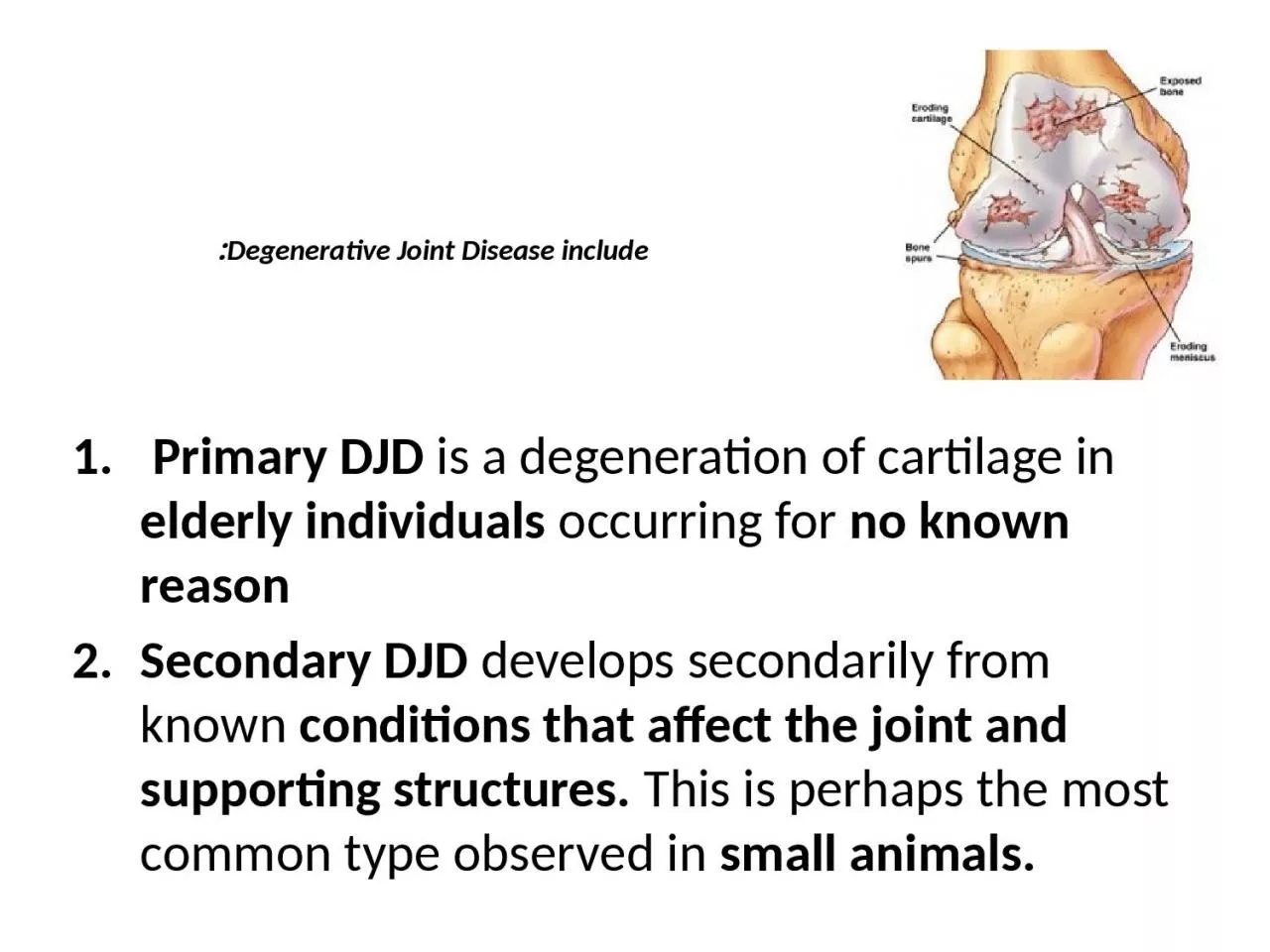
Primary DJD is a degeneration of cartilage in elderly individuals occurring for no known reason Secondary DJD develops secondarily from known conditions that affect
Download Presentation
"Degenerative Joint Disease include:" is the property of its rightful owner. Permission is granted to download and print materials on this website for personal, non-commercial use only, provided you retain all copyright notices. By downloading content from our website, you accept the terms of this agreement.
Presentation Transcript
Transcript not available.