/
Int J Anat Res Int J Anat Res

Int J Anat Res - PDF document

deena
deena . @deena
Follow
342 views
Uploaded On 2022-10-13

Int J Anat Res - PPT Presentation

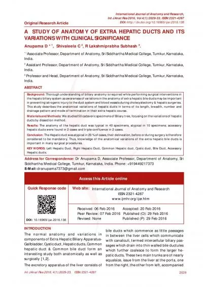
2016 41202933 ISSN 23214287 2029 Original Research Article A STUD Y OF ANA T OMY OF EXTRA HEP A TIC DU C T S AND IT S V ARIA TIONS WITH CLINICAL SIGNIFICANCE Anupama D 1 Shivaleela C ID: 959938

duct hepatic common ducts hepatic duct ducts common cystic extra bile anatomy fig biliary cases 2016 variations study left

Share:

Link:

Embed:

Download Presentation from below link

Download Pdf The PPT/PDF document "Int J Anat Res" is the property of its rightful owner. Permission is granted to download and print the materials on this web site for personal, non-commercial use only, and to display it on your personal computer provided you do not modify the materials and that you retain all copyright notices contained in the materials. By downloading content from our website, you accept the terms of this agreement.


Presentation Transcript

Int J Anat Res 2016, 4(1):2029-33. ISSN 2321-4287 2029 Original Research Article A STUD Y OF ANA T OMY OF EXTRA HEP A TIC DU C T S AND IT S V ARIA TIONS WITH CLINICAL SIGNIFICANCE Anupama D * 1 , Shivaleela C 2 , R Lakshmiprabha Subhash 3 . ABSTRACT Address for Correspondence: Dr Anupama D, Associate Professor, Department of Anatomy, Sri Siddhartha Medical College, Tumkur, Karnataka, India. Phone: +919449217373 E-Mail: dranupama7373@gmail.com Background: Thorough understanding of biliary anatomy is required while performing surgical interventions in the hepato biliary system as awareness of variations in the anatomy of extra hepatic bile ducts may be important in preventing iatrogenic injury to the duct system and blood vessels during cholecystectomy & hepatic surgeries. This study describes the anatomical variations of hepatic ducts in terms of its length, breadth, number and drainage pattern and mode of termination in their extra hepatic course. Materials and Methods: We studied 50 cadaveric specimens of Biliary tree, focusing on the variations of hepatic ducts by dissection method. Results: The anatomy of the hepatic duct was typical in 40 specimens, atypical in 10 specimens; accessory hepatic ducts were found in 2 cases and triple confluence in 2 cases. Conclusion: The Hepatic duct was atypical in 20 % of cases, their delineation, before or during surgery is therefore considered to be mandatory. Thus, knowledge of the anatomical variations of the extra hepatic bile ducts is important in many surgical procedures. KEY WORDS: Left Hepatic Duct, Right Hepatic Duct, Common Hepatic duct, Cystic duct, Bile Duct, Accessory Hepatic ducts. INTRODUCTION International Journal of Anatomy and Research, Int J Anat Res 2016, Vol 4(1):2029-33. ISSN 2321-4287 DOI: http://dx.doi.org/10.16965/ijar.2016.138 Access this Article online Quick Response code Web site: Received: 06 Feb 2016 Accepted: 20 Feb 2016 Peer Review: 07 Feb 2016 Published (O): 29 Feb 2016 Revised: None Published (P): 29 Feb 2016 International Journal of Anatomy and Research ISSN 2321-4287 www.ijmhr.org/ijar.htm DOI: 10.16965/ijar.2016.138 *1 Associate Professor, Department of Anatomy, Sri Siddhartha Medical College, Tumkur, Karnataka, India. 2 Assistant Professor, Department of Anatomy, Sri Siddhartha Medical College, Tumkur, Karnataka, India. 3 Professor and Head, Department of Anatomy, Sri Siddhartha Medical College, Tumkur, Karnataka, India. bile ducts which commence as little passages in between the liver cells which communicate with canaliculi, termed intercellular biliary pas- sages which drain into thin walled bile ductules which further coalesce to form the larger he- patic ducts . These two main trunks are of nearly equalsize, issue from the liver at the porta, one from the right, the other from left, accompanied The normal anatomy and variations in components of Extra Hepatic Biliary Apparatus- Gallbladder, Cystic duct, Hepatic ducts, Common hepatic duct & Common bile duct form an interesting study both anatomically as well a

s surgically [1,2]. The excretory apparatus of the liver consists of Int J Anat Res 2016, 4(1):2029-33. ISSN 2321-4287 2030 Anupama D, Shivaleela C, R Lakshmiprabha Subhash. A STUD Y OF ANA T OMY OF EXTRA HEP A TIC DU C T S AND IT S V ARIA TIONS WITH CLINICAL SIGNIFICANCE. by the hepatic artery and portal vein and unite to form Common Hepatic Duct which is joined by the cystic duct, or the duct of the gallbladder on the right side to form the Common bile duct. Their variations in course and confluence, and the relationship between the structures of the portal pedicle is of much clinical significance. Although congenital anomalies are frequently reported, majority of the cases present during adult life and mimic some acquired conditions [3,4]. The anomaly per se is often associated with calculus formation and duct dilatation [5] . Variations in Cystic & Hepatic ducts are not un- common. In some there can be low or high union of cystic & hepatic ducts or confluence of extra hepatic & cystic ducts. These variations have drawn the attention of Anatomists and Surgeons as there is increased incidence of iatrogenic injuries to these organs or structures in their vicinity which may endan- ger the life of the patient. This is because of failure to recognize at surgeries the unusual anatomical relationships or persistence into adult life of vestigial ductal structures like ac- cessory ducts [6] . Accessory hepatic ducts are normal segmental ducts that join the biliary sys- tem outside the liver instead of within it and are in positions of danger during cholecystec- tomy. Injury to the ducts leads to postoperative bile leakage, shock, peritonitis etc contributing to morbidity and mortality. So the main function of anatomists lie with the detailed study of normal anatomy and variations which give valuable information to the surgeons to conduct surgeries in easiest possible way with good results and to aid in teaching [7,8]. Normal & developmental anatomy: The right and left hepatic ducts emerge from the right and left lobes of liver at the porta hepatis. After a short course, hepatic ducts unite to form Com- mon hepatic duct. The confluence of the right and left hepatic ducts in most of the cases oc- curs outside the liver and in dissections in this area it may be necessary to push liver substance away to display the confluence completely. The common hepatic duct is about 4cm long & 0.4cm in caliber. It is joined by the Cystic duct from the gall bladder on its right side to form the bile duct which runs in the anterior free margin of lesser omentum and it is related behind to the Portal vein & Hepatic artery [9]. Ruge (1908) described and classified the union of Cystic duct & Common hepatic duct to form Common bile duct [10]. Hepatic bud & its Duct system including Gallbladder develop during the 4 th week as a diverticulum from the endoderm of foregut at the ventral border of Duodenum. Hepatic bud further divides into a large cranial portion (Pars hepatica) & a small caudal portion (Pars cystica). Original stalk of hepatic diverticulum forms bile duct, from which a cystic bud develops to form

both Cystic duct & Gallbladder [11]. MATERIALS AND METHODS In the present study a total number of 50 human livers with Gallbladder and its duct system with related ligaments, Duodenum and head of the Pancreas were collected from the cadavers. All the specimens were cleaned thoroughly under running water. The ligaments and other structures were carefully dissected to expose the Cysto hepatic angle. Hepatic ducts and Common hepatic duct were carefully identified. All the specimens were numbered and various measurements of hepatic and common hepatic ducts were taken with the help of measuring tape. Photographs of all the specimens were taken. This study was limited only to the variations in the hepatic duct pattern in its extra hepatic course. RESULTS AND DISCUSSION Table 1: Formation of Common hepatic duct. EHU IHU 1 Ruge 1908 [10] 43 34 9 2 Eisendrath 1920 [7] 100 100 - 3 S G Puente 1944 [3] 3845 3768 77 4 Present study 50 23 27 Sl no Author No of cases Formation of CHD Table 2: Accessory hepatic duct. Sl no Author No of cases No of AHD 1 Brewer 1903 [4] 57 5 2 Osler & Dow 1945 [2] - - 3 Dowdy 1962 [12] 100 15 4 Puente 1944 [3] 3845 - 5 Present study 50 2(Rt) Int J Anat Res 2016, 4(1):2029-33. ISSN 2321-4287 2031 Anupama D, Shivaleela C, R Lakshmiprabha Subhash. A STUD Y OF ANA T OMY OF EXTRA HEP A TIC DU C T S AND IT S V ARIA TIONS WITH CLINICAL SIGNIFICANCE. Table 3: Mode of joining of cystic duct with common hepatic duct. 1 Ruge 1908 [10] 43 100 14 32.5 12 27.9 16 37.2 2 Descomps 1962 [12] 50 100 40 80 6 12 4 8 3 Eisendrath 1918 [5] 100 100 75 75 17 17 8 8 4 Present study 50 100 43 86 1 2 6 12 % No Sl. No Author No of cases Type 1 Angular Type 2 parallel Type 3 Spiral Cases % No % No % Fig.1 Intra hepatic union Fig. 2 Extra hepatic union Fig. 3 Short Common hepatic duct Fig.4 Long Common hepatic duct Fig.5 Low fusion of long cystic duct Fig.6 Confluence of ducts Fig. 7 Cystic duct opening in to RHD Fig. 8 Accessory Hepatic Duct 1 Fig. 9 Accessory Hepatic Duct 2 CHD CD CD RHD LHD CHD CHD CHD CD LHD CD CHD RHD RHD LHD CD LHD RHD CD CHD RHD LHD AHD CD CD AHD LHD RHD CHD Hepatic duct varies in length from extremely short to long type. The length of Common hepatic duct varied from a minimum of 0.7 cm as in Fig 3 to a maximum of 3.5 cm as in Fig 4,with an average of 2.5 cm. The diameter of hepatic duct varied from 0.2 to 0.4 cm with an average of 0.3 cm. Dowdy reported the length of Hepatic duct varying between 0.4 to 6 cm with There is a high incidence of anatomical abnor- mality in the disposition and relations of the extra hepatic bile ducts. Intra hepatic union of ducts was seen in 27 cases as shown in Fig 1 & Extra hepatic union in 23 cases as shown in Fig 2. In the extra hepatic union, the length of Right hepatic and left hepatic ducts were ranging from 0.3- 1.2 & 0.5- 1.2cm. In the present study Int J Anat Res 2016, 4(1):2029-33. ISSN 2321-4287 2032 an average of 4 cm [12] . In Snell’s text book of Anatomy, the length of the hepatic duc

t is said as about 4 cm [13] and in Gray’s text book of anatomy it is said as 3 – 4 cm x 0.5cm [9], with an average of 0.6 cm. Linder and Green (1964) claimed that intra hepatic fusion of Right and left hepatic ducts (to form common hepatic duct) was not unusual, which co insides with our study findings. Yet noted that this contradicted the findings of Johonson and Anson (1952) who maintained that extra hepatic union was the rule in 94% of their cases [14]. In one specimen as in Fig . 5 a low fusion of long Cystic duct with Common hepatic duct, the Cystic duct is invariably longer than normal; it may run along side parallel with the Common hepatic duct before joining it, or twist around the Common hepatic duct, fusing with it either anteriorly or at its left hand border. In this case variable length of Cystic duct always tightly bound to Common hepatic duct before the two actually fuse. Thus, vigorous traction on the Cystic duct may produce marked angulations and tenting of the Common hepatic and bile ducts which may then caught in a clamp. Furthermore, over-diligent efforts meticulously to dissect the ducts apart result in either immediate direct injury to the Common hepatic duct or delayed damage if a part of length of this duct is devascularized. A calculus lying in a long parallel duct may compress the hepatic duct & give rise to obstructive symptoms like those of a calculus in the hepatic duct or common duct. Sometimes a duct may dilate to form a new Gall bladder and be a cause of recurrent symptoms [14]. Sometimes there can be abnormal fusion of Right and Left hepatic ducts with the Cystic duct entering at the confluence as shown in Fig 6. This is also regarded as an abnormally high fusion of the Cystic duct with a (virtually) non existent Common hepatic duct, effectively a trifurcation. In this case either the right or left hepatic ducts may be damaged during Cystic duct ligation. The angulation produced by traction could compromise lumen at the confluence if a tie was placed [14]. Occasionally Cystic duct may enter in to right hepatic duct as in Fig 7 and the danger here is that the right hepatic duct itself may be mistaken for Cystic duct & is tied off and divided where it joins the left hepatic duct and if Chole cysto hepatic duct is damaged inadvertently at operation, bile leakage would contaminate the field [14,15]. Accessory hepatic ducts are normal segmental ducts that join the biliary system outside the liver instead if within it. Most commonly it opens in to Common Hepatic Duct [14]. In our study 2 specimens revealed right accessory hepatic ducts as shown in Fig 8 & 9 opening in to confluence in one and Common Hepatic Duct in another. If such a duct was damaged inadvert- ently at operations, bile leakage would contami- nate the field. If however such an injury went unrecognized at surgeries, post operative bile leakage might produce biliary peritonitis, sub phrenic abcess, biliary fistula and possibly may lead to the development of bile duct stricture [14]. CONCLUSION The Hepatic duct component of Extra hepatic biliary apparatus was studied and following variations were noted do

wn from the post mortem specimens of human by dissection method. By this method Hepatic ducts were displayed. The following conclusion has been made from the observations during the study. Right Hepatic duct varied in measurements from a minimum of 0.3cm to a maximum 1.2 cm with an average of 0.7 cm. Left hepatic duct varied in measurements from a minimum of 0.5 to a maximum of 1.4 cm with an average of 1 cm. Extra hepatic union of these 2 ducts was seen in 23 cases & Intra hepatic union in 27 cases. Right Accessory hepatic ducts were noted in two cases. The variations in the duct could lead to damage to it and also to the ducts and vessels in its vicinity. As the area is remarkably variant & with the advent of endoscopic surgeries, sound knowledge regarding the area is very essential. LHD - Left Hepatic Duct RHD - Right Hepatic Duct CHD - Common Hepatic duct CD - Cystic duct BD - Bile Duct AHD - Accssory hepatic duct ABBREVIATIONS Anupama D, Shivaleela C, R Lakshmiprabha Subhash. A STUD Y OF ANA T OMY OF EXTRA HEP A TIC DU C T S AND IT S V ARIA TIONS WITH CLINICAL SIGNIFICANCE. Int J Anat Res 2016, 4(1):2029-33. ISSN 2321-4287 2033 Anupama D, Shivaleela C, R Lakshmiprabha Subhash. A STUD Y OF ANA T OMY OF EXTRA HEP A TIC DU C T S AND IT S V ARIA TIONS WITH CLINICAL SIGNIFICANCE. Conflicts of Interests: None REFERENCES [1]. Hicken. Anatomic Variations of Extrahepatic Biliary Apparatus. Surg; Obst; Gynae1954;10:577-84. [2]. Osler.G .& Dow.R. Variations and anomalies of the biliary duct system and its associated blood supply.WestJ.Surg1945;53:316-21. [3]. Sandrini. Congenital abnormalities of bile passage.Arch of surgery1944;(1):262-66 . [4]. Brewer.G.E. Anatomy of the Gallbladder region. Medi- cal news.1903;82:821-23. [5]. Agvey.I.Rogeus. Congenital absence of Gallbladder with Choledocholithiasis. Gastroenterol- ogy.1965;48:4. [6]. Moosman. Anomalies of Biliary tract System. Surg, Obst, Gynae.1970; 82: 655-62. [7]. Eisendrath. Surg, Obst, Gynae. Operative Injury to Common hepatic & Bile Ducts. Surg, Obst, Gynae 1920;31:1-18. [8]. Lurje et al. Topography of Extra Hepatic Biliary pas- sage. Annals of Surgery.1937; 105:161-168. [9]. Henry gray: In Gray’s Anatomy. 39 th ed.Edinburgh: Elsevier.1995; p 1228-30. [10].Ruge.E. Anomalies of Bileducts .Arch.F. Klin. Chi .1908;77:47-78. [11]. T.W.Saddler In:Langman’sMedical Embryology.10 th ed.London:Lippincott Williams Wilkins;2006.p213- 17. [12]. Dowdy et al. Surgical Anatomy of Pancreatic Biliary System. Surgery.1962;108(6):p 233-34. [13].Richard Snell. Snell’s Text Book of Anatomy 5 th ed.1995. p219-20. [14]. E.A.Benson, R.E.Page. A practical reappraisal of the anatomy of the extra hepatic bileducts and arteries.British Journal of Surgery.1976;63:853-860. [15]. Daniel.N.Eisendrath. Anomalies of Bile ducts and blood vessels. Journal of American Medical Asso- ciation.1918;71:864. How to cite this article : Anupama D, Shivaleela C, R Lakshmiprabha Subhash. A STUDY OF ANA T OMY OF EXTRA HEP A TIC DU C T S AND IT S V ARIA TIONS WITH CLINICAL SIGNIFICANCE. Int J Anat Res 2016;4(1):2029- 2033. DOI: 10.16965/ijar.2016.1