/
Skin  Care   and  Advanced Skin Skin  Care   and  Advanced Skin

Skin Care and Advanced Skin - PowerPoint Presentation

eatsyouc
eatsyouc . @eatsyouc
Follow
343 views
Uploaded On 2020-06-18

Skin Care and Advanced Skin - PPT Presentation

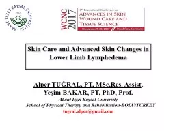
Changes in Lower Limb Lymphedema Alper TUĞRAL PT MScRes Assist Yeşim BAKAR PT PhD Prof Abant Izzet Baysal University School of Physical Therapy ID: 781193

amp skin infection care skin amp care infection wound chronic integration patients moisture cellulitis lymphoedema journal treatment folds due

Share:

Link:

Embed:

Download Presentation from below link

Download The PPT/PDF document "Skin Care and Advanced Skin" is the property of its rightful owner. Permission is granted to download and print the materials on this web site for personal, non-commercial use only, and to display it on your personal computer provided you do not modify the materials and that you retain all copyright notices contained in the materials. By downloading content from our website, you accept the terms of this agreement.


Presentation Transcript

Slide1

Skin Care and Advanced Skin Changes in Lower Limb Lymphedema

Alper TUĞRAL, PT,

MSc,Res. Assist, Yeşim BAKAR, PT, PhD, Prof.Abant Izzet Baysal University School of Physical Therapy and Rehabilitation-BOLU/TURKEYtugral.alper@gmail.com

Slide2

Presentation PlanNormal Skin StructureSkin changes and problems specific to LymphedemaSkin care in Lymphedema

Slide3

Normal SkinProtective barrier functionPrimary determiner of maintaining natural moisture balance.

Slide4

Importance of skin moisture

Slide5

Skin changes in LymphedemaStructural fibroproliferative tissue changes-Activation of Fibrin and WBCFibroproliferation Epidermal, dermal and soft Tissue thickening.Structural

degeneration in capillary

 IneffectivityResearchers also pointed out anormal expression of inflamatory cytokines, adhesion molecules and growth factors.

Slide6

Background PhysiologyProlonged and increased interstitial pressureSkin changes. Changes in perfusion and nutrition. Underlying reason, changes in adipose

and fibrous

tissue in basic. (Olzewski, 2003)Diminished elasticity, natural moisture balance is lost. Advanced skin changes Hyperkeratosis, Papillamatosis etc.

Slide7

ImpactsAdvanced skin changes cause patients to be more vulnerable to infection in addition to corruption of barrier function of skin.Moffatt (2003): Incidence of cellulitis infection in patients with chronic edema is 29%.25% of them or above, hospitalization is required!

Moffatt, C. J.,

Franks, P. J., Doherty, D. C., Williams, A. F., Badger, C., Jeffs, E., ... & Mortimer, P. S. (2003).Lymphoedema: an underestimated health problem. Qjm, 96(10), 731-738. ISO 690

Slide8

Diagnosing and intervention to skin problems in early stage prevent the cycle to get worse and improve patients’ quality of life. Besides, improvement in progression of disease and reducing health care costs could be achieved by early intervention.

Hofman, D. (2010). Managing ulceration caused by oedema. Wounds Essentials, 

5, 80-6.

Slide9

Skin changes and problems specific to LymphedemaCellulitis (Erysipelas)HiperkeratosisPapillamatosisLymphorrhoea (Lymph Fistula)Fungal infectionsFolliculitisUlcerations Skin folds

Slide10

Cellulitis (Erysipelas)Inflamatory reaction which includes tissues in skin and sub-skinTypical inflamation signs (+). β-hemolitic streptococcus.Group A streptococcus according to some researchersMicrobiologists: Staphylococcus Aureus in some

patients.Mortimer, P. (2000). 9 Acute inflammatory episodes. Lymphoedema, 130.

Al-Niaimi, F., & Cox, N. (2009). Cellulitis and lymphoedema: a vicious cycle. Journal of Lymphoedema, 4(2), 3-42.Chira, S., & Miller, L. G. (2010). Staphylococcus aureus is the most common identified cause of cellulitis: a systematic review. Epidemiology and infection, 138(03), 313-317.

Slide11

Cellulitis (Erysipelas)Strong correlation with LE together with reduced immune system function.Lower extremity is the most affected area96 M £ each year according to the NHS7.1 days hospitalization in average!Dupuy, A., Benchikhi, H., Roujeau, J. C., Bernard, P., Vaillant, L., Chosidow, O., ... &

Bastuji-Garin, S. (1999). Risk factors for erysipelas

of the leg (cellulitis): case-control study. Bmj, 318(7198), 1591-1594.Cox, N. H., Colver, G. B., & Paterson, W. D. (1998). Management and morbidity of cellulitis of the leg. Journal of the Royal Society of Medicine, 91(12), 634-637.Halpern, J., Holder, R., & Langford, N. J. (2008). Ethnicity and other risk factors for acute lower limb cellulitis: a UK‐based prospective case–control study. British Journal of Dermatology, 158(6), 1288-1292.UK Dermatology Clinical Trials Network's PATCH Trial Team. (2012). Prophylactic antibiotics for the prevention of cellulitis (erysipelas) of the leg: results of the UK Dermatology Clinical Trials Network’s PATCH II trial. The British journal of dermatology, 166(1), 169.

Slide12

Integration to treatmentIn case of acute erysipelas Broad spectrum antibiotics (14-21 days).Topical antibiotics

.MLD ve CDT contraindicates

.

Slide13

Ko et al: CDT reduces frequency of infection attacksKo, D. S., Lerner, R., Klose, G., & Cosimi, A. B. (1998). Effective treatment of lymphedema of the extremities. Archives of Surgery, 133(4), 452-458.CDT

carries a primary

importance regarding the prevention of recurrence and lymphatic improvement after stabilization is maintained

Slide14

HyperkeratosisExcessice thickening of external layer of skin (SC) (ILF,2012).Epidermal hyperplasia due to lymph stasisPhysiology: Hyperproliferative keratinocytes.Characterized by Brown-gray color, fissures and fractures (Day

and Hayes, 2008).Fissures cause

bacterial and fungal colonization Infection!

Slide15

Slide16

Slide17

There is no exact standardized approach. (Young, 2010).Bacterial colonization+ fungal infections can be exist, therefore those all resulted recurrent infections and corruption of skin integrity by creating a cyclic

cycle (Day and Hayes, 2008).

Maintaning daily hygiene and integration of self-care are primarily important in the treatment (Whitaker 2012, Pidock and Jones,2013).Young T (2011) EWMA poster. A national survey of the nursingpractice of the treatment of hyperkeratosis associated with venoushypertension. EWMA Conference. Bruges. BelgiumDay, J., & Hayes, W. (2008). Body image and leg ulceration. Leg Ulcers and Problems of the Lower Limb: An Holistic Approach.Whitaker, J. (2012). Self-management in combating chronic skin disorders. Journal of Lymphoedema, 7(1), 46-50.PIDCOCK, L., & JONES, H. (2013). Use of a monofilament fibre debridement pad to treat chronic

oedema-related

hyperkeratosis

Wounds

UK, 9(3).

Slide18

What can be done?Integration to TreatmentElimination of hyperkeratotic plaques Whitaker, J. (2012). Self-management in combating chronic skin disorders. Journal of Lymphoedema, 7(1), 46-50Using monofilament debridement pads (MDP). NICE Medical Technologies Guidance {MTG17] (2014) The Debrisoft monofilament debridement pad for use in acute or chronic wounds. Available from www.nice.org.uk/guidance/ MTG17MDP is effective in clinical usage. Using this in LE patients is also

effective. Gray, D., Cooper, P., Russell, F., & Stringfellow, S. (2011). Assessing the clinical performance of a new selective mechanical wound debridement product. 

Wounds UK, 7(3), 42-6. Bahr, S., Mustafi, N., Hättig, P., Piatkowski, A., Mosti, G., Reimann, K., ... & Abb42-8. McGrath, A. (2013). The management of a patient with chronic oedema: a case study. British journal of community nursing.ritti, F. (2011). Clinical efficacy of a new monofilament fibre-containing wound debridemen.t product. J Wound Care, 20(5), 2. PIDCOCK, L., & JONES, H. (2013). Use of a monofilament fibre debridement pad to treat chronic oedema-related hyperkeratosis. Wounds UK, 9(3).

Slide19

Proper MoisturizerWater based moisturizers are not effective regarding moisturizing in the treatment of hyperkeratosis.Moncrieff, G., Cork, M., Lawton, S., Kokiet, S., Daly, C., & Clark, C. (2013). Use of emollients in dry‐skin conditions: consensus statement. Clinical and experimental dermatology, 38(3), 231-238.250-600 gr/week is convenient in adultsErsser, S., Maguire, S., Nicol, N., Penzer, R., & Peters, J. (2007). Best practice in emollient

therapy: a statement for healthcare

professionals. Dermatology Nursing, 6(4).Patient compliance is the most important parameter regarding moisturizing and skin care protocolCork, M. J., & Danby, S. (2009). Skin barrier breakdown: a renaissance in emollient therapy. British Journal of Nursing, 18(14).

Slide20

PapillamatosisBenign skin growths characterized by epithelial neoplasms.Villous/fibrous vascular structures.Tends to hyperkeratosis.Vulnerable to mechanical traumas and can be easily bleed due to vascular background.

Slide21

Slide22

Slide23

Slide24

Slide25

Slide26

Slide27

Integration to TreamentSkin care and protection of natural moisture balance of skin with urea based creamsThey should be supported during the application of compression bandageShould be protected againts mechanical traumas: Infection risk!Surgical

debridement? Immune

inefficacy must be taken into account

Slide28

Lymphorrhea (Lymph Fistula)Frequent in sides where interstitial pressure increased and reduced lymphatic transportLymph cyst Trauma Lymph Fistula Leakage of serum fluid

It might

be spontaneous in primary lymphedema (Cardone-Gaines and Khachemoune, 2013).Infection risk increased due to contact between acidic fluid and intact skin (+++).

Slide29

Wet Legs-Odour-Tend to be infected

Slide30

Integration to TreatmentFluid absorbent wound padsCompression bandage with fluid absorbent wound pads.Cook (2011): Attention to sub-bandage pressure. (Due to the capacity of absorbent

pads)Potassium permanganate: Effective in

exudative inflamatory degenerations.High protein concentration: Inflamation!Cook, L. (2011). Effect of super-absorbent dressings on compression sub-bandage pressure. British Journal of Community Nursing, 16(3), 38.

Slide31

Absorbent PadsHydrophobicity; provides adhering bacteries and other infectious agents to wound pads, remove them from wound by preventing bacterial colonization in an infected wound.

Slide32

Key Features of Wound Pads

Slide33

Fungal InfectionsFungal growth in dead keratin (+++)Accumulation of keratin is frequent in LEProtein: Fasilitator of the growth of fungal infectionsHygiene problems between skin folds, increased

temperature, friction

between skin folds: Frequent infection!Recurrent infections: Affect the treatment negatively.Especially in interdigital areas!

Slide34

Integration to TreatmentAntifungal topical applicationsSupporting the gaps between skin folds (Reducing friction stress)Maintaining hygieneCompression bandage with topical antifungal applications.Contact exposure!

Slide35

FolliculitisInflamation of hair follicleCan include fungal/ bacterial componentFrequent in LE: Folliculitis due to mechanical irritation

Slide36

Integration to TreatmentMaintaining hygieneDetermination of factors which cause irritationEspecially in males, moisturizer should be applied to downward direction due to the hair growth.Infected area

should be supported with anti-microbial

agents.

Slide37

UlcerationDysfunctional immune systemProlonged tissue hypoxiaConcomitants(Infection, Trauma)In lower limb LE:Ulcerations due to vascular insufficiency.Increased

interstitial

fluid Increased diffusion distance(O2 Dermal capillaries)

Slide38

Slide39

Important! Determination of EtiologyPhlebolymphedema Ulceration with venous background

Slide40

Compression EffectsHealed ulcerations only with compression treatment 70%Improves peripheric pump activationIncreases venous blood flow rateReduces venous reflux.Reduces venous blood volume.Increases lymphatic drainage, reduces

edema

Important: Compression treatment should be combined with active movement. Efficacy can be increased more by this way.

Slide41

Venous Stats1.5-2 M patients with leg ulcer. 200.000 new cases per annum70% of them originating from CVIUlcer manifested 1 year ago at least 50% of patientsRecurrence rate after healing 60-90%

(Joachim Dissemond 2007:

Ulcus-cruris doğuşu,tanılama,tedavi. UNI-MED araştırma)

Slide42

Venous Stats5% of patients aged over 80 years or aboveTotal treatment costs: 1-1.5 billion €Healing rates after 3 months: %66-90In Ulcus Cruris Venosum:-30% of patients heal totaly

-Recurrence rate: 70%. Why?

Slide43

Slide44

Integration to TreatmentCause of ulceration?! Infection?, Trauma?  Should be determined.Absorbent wound pads+

compression.Maintaining

tissue sterilization with anti-inflamatory agents (Topical antimicrobial applications)Infection control!

Slide45

Skin FoldsIncreased edemaGravity effectSkin elasticityBetween folds: Infection risk MacerationInfection Edema!

Slide46

Slide47

Slide48

Slide49

Integration to TreatmentMaintaining the optimal hygiene betwen skin folds:-Anti-bacterial, anti-fungal topical agentsIntegration to CDT should be done by supporting folds with proper filling

materials (equal

pressure distribution)Maintaining moisture balance (Skin tend to be more fragile in skin folds).

Slide50

Skin Care in LymphedemaRegardless of etiology, in all chronic edema skin care is crucial!Providing optimal moisture balance: Primary protection mechanism to infections and skin integrity

Timmons, J., & Bianchi, J. (2008). Disease progression in venous and lymphovenous disease: the need for early identification and management. 

Wounds UK, 4(3), 59-71.

Slide51

Why is Skin Care important?Providing the lipid layer on skin and hidrationMoffatt (2006): Protecting the skin from bacteria and other infectious agents, adherence to achievements of treatment: Skin Care!Skin care: Improvement of barrier function

!Stephen-Haynes, J. (2007). Skin care in chronic oedema. 

Wounds UK, 3(2 Suppl), 1-40.Moffatt, C. J. (2006). Skin care management for patients with lymphoedema. Wound Essentials, 1, 172-4.

Slide52

Skin care and maintaining optimal moisture balance are more advantageous than medications regarding the prevention of infections (Badger, 2004).Badger, C., Preston, N., Seers, K., & Mortimer, P. (2004). Antibiotics/anti‐inflammatories for reducing acute

inflammatory episodes in lymphoedema of

the limbs. The Cochrane Library.

Slide53

MoisturizersEach moisturizer is not the same Working principle: Moisturizing/ epidermal dermal penetration and moisture (urea,glycerin content).Epidermal

moisture:

Could be exfoliative/ anti inflamatory. BDNG (2012)British Dermatological Nursing Group (BDNG) (2012) Best Practice in Emollient Therapy. A statement for healthcare professionals. Dermatol Nurs 11(4)

Slide54

MoisturizerProper choice Without any perfume or smell.Oil based moisturizer?  Lymphedema?Dry skin paraffin based

(50/50) Moffatt, C. J. (2006). Skin care management for patients with lymphoedema. Wound Essentials, 

1, 172-4.Skin condition and hidrationPersonal choiceCosmetic acceptability proper pH interval.

Slide55

ImportantPatient education!Self monitorization of skinRecognizing infection signsImportance of skin careDownward application to hair growthChoice of proper products

Slide56