/
S K Yoon and S H Rha15 of RCC including those previously referre S K Yoon and S H Rha15 of RCC including those previously referre

S K Yoon and S H Rha15 of RCC including those previously referre - PDF document

eddey
eddey . @eddey
Follow
343 views
Uploaded On 2022-09-06

S K Yoon and S H Rha15 of RCC including those previously referre - PPT Presentation

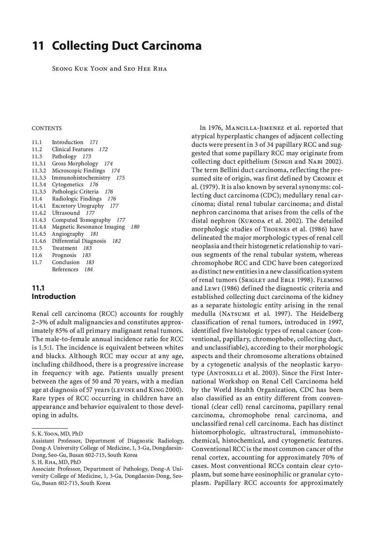
Fig 111 Normal histologic anatomy of the kidney Collecting Duct Carcinoma family history of associated malignancies including colon pancreas lung ovary and uterus malignancies Kuroda et al 2 ID: 951579

collecting renal carcinoma duct renal collecting duct carcinoma tumor fig cdc rcc patients shows kidney tumors 2002 year medullary

Share:

Link:

Embed:

Download Presentation from below link

Download Pdf The PPT/PDF document "S K Yoon and S H Rha15 of RCC including ..." is the property of its rightful owner. Permission is granted to download and print the materials on this web site for personal, non-commercial use only, and to display it on your personal computer provided you do not modify the materials and that you retain all copyright notices contained in the materials. By downloading content from our website, you accept the terms of this agreement.


Presentation Transcript

S. K. Yoon and S. H. Rha15% of RCC, including those previously referred to as chromophil RCC. Chromophobe RCC accounts for approximately 5% of cases of RCC. The cytoplasm contains numerous microvesicles that appear blue with Hales colloidal iron stain. Unclassified RCC is reserved for cases that do not fulfill the criteria for the cancers described previously. These tumors are morphologically and genetically variable and are often high grade. Less than 2% of renal carcinomas are unclassified (Bostwick and Eble 1999The collecting duct is embryologically derived from the ureteric bud, whereas the remainder of the kidney develops from metanephric blastema. The ureteric bud also differentiates into ureter, renal pelvis, and calyces. The renal collecting ducts extend from the cortex to the tips of the renal papillae Srigley and Eble 1998). Collecting duct carcinoma, or Bellini duct carcinoma, is a very rare, aggressive renal neoplasm that arises from the distal segment of the collecting ducts of Bellini in the renal medullary pyramids (Fig. 11.1), unlike the much more common variants of RCC which arise from the convoluted tubules (Sironi et al. 20

03). Collecting duct carci-noma originates in the medullar collecting duct, which arises from the mesonephron. In contrast, the tubular structures of the kidney, which can be a point of origin for RCC, originate in the metanephric blastema (Auguet et al. 2000). Recognition of CDC remains somewhat controversial, and the tumor is typically described as containing irregular channels lined by highly atypical epithelium, often with a hob-nail cell appearance (Bostwick and Eble 1999). Col-lecting duct carcinoma is almost invariably centered in the renal medulla with or without extension into the renal cortex, renal pelvis, or hilar soft tissue. Its hypothesized origin from the collecting duct epithe-lium is supported by the presence of the collecting duct epithelium dysplasia far from the main tumor and reactivity of tumor cells with antibodies to high molecular weight cytokeratin and Ulex europaeusagglutinin-I (UEA-I; Gong et al. 2003The most common neoplasms arising from the collecting ducts are renal oncocytoma and chro-mophobe RCC, which appear to arise in the cortex from the intercalated cells of the proximal connect-ing segment of the collecti

ng ducts. Collecting duct carcinoma is a much less common neoplasm that displays a tubulopapillary architecture, arises in the renal medulla (and later invades the renal cortex), and demonstrates infiltrating tubules with an associated desmoplastic reaction (Milowski et al2002). The microscopic appearance of the tumor is variable; thus, diagnosis based on histologic criteria alone cannot be accurate, and immunohistochemi-cal staining is needed to demonstrate the origin of the tumor (Kirkali et al. 1996). Because most CDC cases are published as case reports, it is difficult to characterize this entity.In this chapter, we review the natural history, clinical course, pathologic and radiologic findings, treatment, and prognosis of typical CDCs.11.2 Clinical FeaturesCollecting duct carcinoma is a rare variant of renal carcinoma associated with an aggressive course and an extremely poor prognosis. It is considered one of the most aggressive carcinomas of the renal tubular epithelium: 35…40% of patients with distant metas-tases and two-thirds of all patients are dead within 2 years of diagnosis (Gallob et al. 2001). Collecting duct carcinoma account

for approximately 0.4…2.6% of all renal neoplasms. With regard to the incidence of CDC, Rumpelt et al. (1991) found six CDC (0.4%) among 1400 consecutive RCC. Most cases reported previously had a tendency to early dissemination and a fatal clinical course. Patients tend to be younger than those with classical RCC (median age at diag-nosis of 43 years) and white (Milowski et al. 2002; Natsume et al. 1997). More men than women (ratio 2:1) are affected. There is no predominant laterality of the affected side. Patients with CDC usually have a Fig. 11.1. Normal histologic anatomy of the kidney. Collecting Duct Carcinoma family history of associated malignancies, including colon, pancreas, lung, ovary, and uterus malignancies Kuroda et al. 2002). Clinical manifestations of CDC, similar to those of RCC, are hematuria, flank pain, and palpable mass. Constitutional symptoms, such as fever, anorexia, and weight loss, are also common, but no particular paraneoplastic syndrome has been reported (Auguet et al. 2000; Pickhardt 1999).The tumor typically follows an aggressive clinical course with most patients showing evidence of meta-static disease at prese

ntation. The most common site of metastatic disease is the lymph nodes, including the cervical or supraclavicular lymph nodes and vis-ceral metastases. Nodal involvement was reported in 77.8% and lung metastases in 44.4% of patients Peyromaure et al. 2002). These findings are simi-lar to those of Davis et al. (1995), who reported nodal involvement and lung metastasis rates of 78 and 27%, respectively. In their study tumors were also metastatic to the adrenal gland in 24% of cases and to the liver in 18%. In contrast to RCC, blastic bone metastases occur more frequently than lytic bone metastases in CDC. This rapid metastatic spread may be due to the tumors central location, close to the renal hilus (Milowski et al. 2002; Pey-romaure et al. 2002). The growth pattern of CDC seems to be quite rapid, which would explain why it is almost always detected clinically (90% of our patients), especially when tumors are large (mean 94 mm). This is unlike other renal malignancies, which are increasingly more frequently discovered fortuitously (Mejean et al. 2003).The association of CDC with sickle cell trait (SCT) has been noted by others. In one study 44

.4% of patients were black and 55.6% had SCT. All black patients had SCT. In the series of Davis et al. (1995), which included 33 patients with CDC, all except 1 were black and had SCT (Peyromaure et al. 2002).11.3 PathologyThere are five distinct subtypes of RCC: conven-tional (clear cell); papillary; chromophobe; col-lecting duct subtypes; and unclassified RCC. Each subtype has distinct histomorphologic, ultrastruc-tural, immunohistochemical, histochemical, and cytogenetic features (Gong et al. 2003). A modern classification of renal epithelial neoplasms has been proposed that takes into account both the morpho-logic and genetic findings.Collecting duct carcinoma is an adenocarcinoma consisting of two components. The first component is the main tumor mass, which is papillary with a fibrovascular core, whereas the second, invasive part is composed of glandular elements associated with marked desmoplastic reaction. The tumor cells are columnar, and show variable degrees of pleo-morphism (Nguyen and Schumann 1997).Several pathologic criteria define CDC. It has tubu-lopapillary architecture, arises in the renal medulla, and contains atypical hyp

erplastic changes in adja-cent collecting tubules. Staining for high molecu-lar weight cytokeratins and UEA-I lectin provides strong support for an origin in the collecting duct of the distal nephron. Some cytogenetic abnormali-ties have been identified, including loss of hetero-zygosity of 8p and 13q, which suggests that tumor suppressor genes on 8p and 13q may be involved in the pathogenesis of this variety of cancer (Peyro-maure et al. 2002). The tumor can also have promi-nent papillary or sarcomatoid features, may contain mucin, and can resemble urothelial carcinoma with glandular differentiation or adenocarcinoma aris-ing from the urothelium of the renal pelvis (Gallobet al. 2001). The collecting duct is embryologically derived from the ureteric bud, which explains the urothelial features of some cases of CDC. The patho-logic and immunohistochemical description as well as the cytogenetic abnormalities support the belief that CDC is more similar to urothelial carcinoma than to clear cell RCC (Milowski et al. 2002).Two other neoplasms considered to originate from the collecting ducts, renal medullary carcinoma Davis et al. 1995) and low-gra

de CDC (MacLen-nan et al. 1997), have been described. Renal medul-lary carcinoma arises in young black patients with SCT, and has a reticular, yolk sac tumor-like, or adenoid cystic morphology. There is some overlap in histologic appearance between typical CDC and renal medullary carcinoma, with the latter seem-ing to form the aggressive end of the CDC spectrum Srigley and Eble 1998). All patients with this tumor died of the disease; mean duration post-surgery was 15 months. Low-grade CDC presents a grossly solid or cystic, well-circumscribed, macroscopic appear-ance and is characterized by tubular formation of tumor cells with a hobnail cell appearance and low nuclear grade. This tumor seems to have a better prognosis than classic CDC (Fukunaga 1999). Col-lecting duct carcinoma appears to belong to a broad spectrum of distal CDC tumors with different mor-phologic traits and prognoses, which emphasizes the difficulty of diagnosing this condition. Collecting Duct Carcinoma fat. The tumor biologic behavior is mostly aggressive with a high rate of local, lymphatic, and hematog-enous spreading at diagnosis and a poor long-term prognosis. The tumor

consists of an intermin-gling of clear, eosinophilic, or basophilic cells and polymorphic nuclei, often of hobnail appearance (Fig. 11.8); these are diagnostically helpful because hobnail cells are not seen in clear cell, papillary, or chromophobe RCC (Srigley and Eble 1998). Most CDCs have high-grade nuclear features. The plasma is lesser in amount and lacks the charac-teristic clear, granular and spindle cell types seen with RCC in which the nuclei are vesicular and firm and differentiation from metastatic lesions might be difficult, especially from ovarian and pancreatic origin tumors. The presence of dysplastic features in the epithelium of collecting ducts near a tumor has been taken as evidence the tumor originated in the epithelium of the collecting ducts (Fig. 11.9; Antonelli et al. 2003; Singh and Nabi 2002). Col-lecting duct carcinoma with sarcomatoid transfor-mation has been noted (Aita et al. 2003; Kurodaet al. 2002).11.3.3 ImmunohistochemistryHistopathologic differentiation of CDC from RCC is possible only by immunohistochemistry. In lectin histochemistry, Ulex europaeus agglutinin-I is com-monly reactive. Peanut lectin is also o

ften positive. Fig. 11.4 Collecting duct carcinoma in a 45-year-old man. Pho-tomicrograph shows a tubular growth pattern (hematoxylin and eosin stain; original magni“ cation, Fig. 11.5. Collecting duct carcinoma in a 45-year-old man. Photomicrograph shows tubulopapillary growth pattern (hematoxylin and eosin stain; original magni“ cation, Fig. 11.6. Collecting duct carcinoma in a 45-year-old man. Photomicrograph shows an in“ ltrative growth with prominent desmoplastic reaction (hematoxylin and eosin stain; original magni“ cation, Fig. 11.7. Collecting duct carcinoma in a 45-year-old man. Photomicrograph of the collecting duct carcinoma depicts an intratumoral cystic area (hematoxylin and eosin stain; origi-nal magni“ cation, Collecting Duct Carcinoma however, are often large at presentation and also have an expansile appearance and exophytic component that cannot be reliably distinguished from the more common cortical RCC. It cannot always be deter-mined from radiologic images whether a renal tumor in a central location originated in the collecting ducts of the renal medulla.Radionuclide studies seem to show uptake of Ga-67 in CDC, indica

ting that Ga-67 scintigraphy may be helpful for identifying this type of cancer preop-eratively (Peyromaure et al. 2002).11.4.1 Excretory UrographyDistortion of the intrarenal collecting system, with calyceal displacement and associated filling defects is observed. Medial displacement of the ureter can be seen. Focally prolonged cortical retention of con-trast material may be seen on delayed film in the region that supports the medullary portion involved by tumor.11.4.2 UltrasoundCollecting duct carcinoma appears as a solid tumor with variable echogenicity compared with normal renal parenchyma, although most lesions are reported to be hyperechoic (Fig. 11.10). A hypoechoic rim is not identified at the tumor…parenchyma interface, indicating the absence of a pseudocapsule with this infiltrative neoplasm. Anechoic cystic areas are noted, representing either intratumoral cysts or cystic necrosis.11.4.3 Computed TomographyCollecting duct carcinoma generally shows a hetero-geneous solid renal mass arising from the central kidney region with extension to the renal sinus. The mean size of the tumors is 7.7 cm (range 1.5…19 cm). Most tumors exhibit inv

olvement of the renal medulla (Fig. 11.11). Recognition of a medullary origin can be difficult in larger tumors. Replace-ment of the renal sinus fat or protrusion into the renal pelvis is present in many cases (Fig. 11.12). Occasionally, CDC reveals an exophytic appearance without involvement of the renal sinus (Fig. 11.13). Table 11.1. Diagnostic criteria for collecting duct carcinoma Major criteriaMinor criteriaLocation in a medullary pyramid (small tumors)Central location (large tumors)Typical histology with irregular tubular architecture, desmopla-sia, and high gradePapillary architecture with wide, fibrous stalks, and desmo-plastic stromaReactive with antibodies to high-molecular weight cytokeratinInflammatory stroma with numerous granulocytesReactive with Ulex europaeus lectinExtensive renal, extrarenal, and vascular infiltrationAbsence of urothelial carcinoma Fig. 11.10. Collecting duct carcinoma in a 45-year-old man. Longitudinal US image shows a poorly de“ ned, hypoechoic arrow) at the lower pole of the right kidney. Fig. 11.11. Collecting duct carcinoma in a 55-year-old woman. Axial contrast-enhanced CT scan shows a poorly marginated

enhancing mass in the renal medulla of the right kidney. Collecting Duct Carcinoma Fig. 11.15a-c. Collecting duct carcinomas are classi“ ed into three types according to the main location and the pattern of tumor growth ( tumor). Medullary involvement only. Medullary involvement with cortical extension. Medullary involvement with renal sinus extension. Fig. 11.16a,b. Collecting duct carcinoma in a 62-year-old man. Axial contrast-enhanced CT scan shows left kidney ill-de“ ned tumor with in“ ltrative growth pattern. Gross specimen shows an in“ ltrative growth with poor demarcation in the renal medulla. Fig. 11.17a,b. Collecting duct carcinoma in a 45-year-old man. Axial contrast-enhanced CT scan shows an expansile growth in the right kidney with bulging tumor margins. Gross specimen shows an ill-de“ ned protruding mass at the lower pole. S. K. Yoon and S. H. Rhaimal or moderate enhancement (Figs. 11.20, 11.21). Gross extension to perinephric fat and vascular inva-sion are sometimes present (Fig. 11.22). Calcification is rare, unlike in conventional RCC (Fig. 11.23). A cystic component that resembles the more conven-tional cortical RCC,

suggesting either intratumoral cyst or cystic necrosis, is frequently noted within the tumors (Fig. 11.24).Many patients with CDC already have invasion into other sites, lymphadenopathy, or metastasis, when diagnosis is established. About 35-40% of patients have metastases at presentation. Regional lymph nodes (60…80%) (Fig. 11.25), lung (Fig. 11.26) or adrenal glands (25%), bone, and liver are common metastatic sites. Lymph node metastasis and inferior vena caval thrombosis are also present. Metastases to bone appear osteolytic or osteoblastic appearance (Fig. 11.27; Kuroda et al. 2002).In summary, medullary location, involvement of the renal sinus, infiltrative growth, preservation of renal contour, and weak enhancement are CT find-ings most commonly seen in patients with CDC of the kidney.11.4.4 Magnetic Resonance ImagingCollecting duct carcinoma appears isointense to normal renal parenchyma on T1-weighted spin-echo images and hypointense to normal renal parenchyma on T2-weighted spin-echo images. As with CT, weak enhancement is noted on contrast-enhanced T1-weighted image (Fig. 11.28). The lack of a visible marginal hypointense rim indicat

es that there is no pseudocapsule. In general, RCC appears Fig. 11.18. Collecting duct carcinoma in a 62-year-old woman. Axial contrast-enhanced CT scan reveals a heterogeneous mass in the right kidney (arrow) with preservation of the renal outer contour. Fig. 11.19. Collecting duct carcinoma in a 65-year-old man. Axial contrast-enhanced CT scan shows the left kidney tumor appears exophytic as collecting duct carcinoma enlarges.Fig. 11.20a,b. Collecting duct car-cinoma in a 49-year-old man. Axial unenhanced CT scan shows a hypodense mass (arrow) at the left renal medulla. Axial contrast-enhanced CT scan shows minimal enhancement of the tumor. ba Collecting Duct Carcinoma Sironi M, Delpiano C, Claren R et al. (2003) New cytological findings on fine-needle aspiration on renal collecting duct carcinoma. Diagn Cytopathol 29:239…240Srigley JR, Eble JN (1998) Collecting duct carcinoma of kidney. Semin Diagn Pathol 15:54…67Thoenes W, Störkel S, Rumpelt HJ (1986) Histopathology and classification of renal cell tumors (adenomas, oncocytomas and carcinomas). The basic cytological and histopathologi-cal elements and their use for diagnostics. Pathol Re