/
A B C Centromere DNA sequence A B C Centromere DNA sequence

A B C Centromere DNA sequence - PowerPoint Presentation

edolie
edolie . @edolie
Follow
64 views
Uploaded On 2024-01-29

A B C Centromere DNA sequence - PPT Presentation

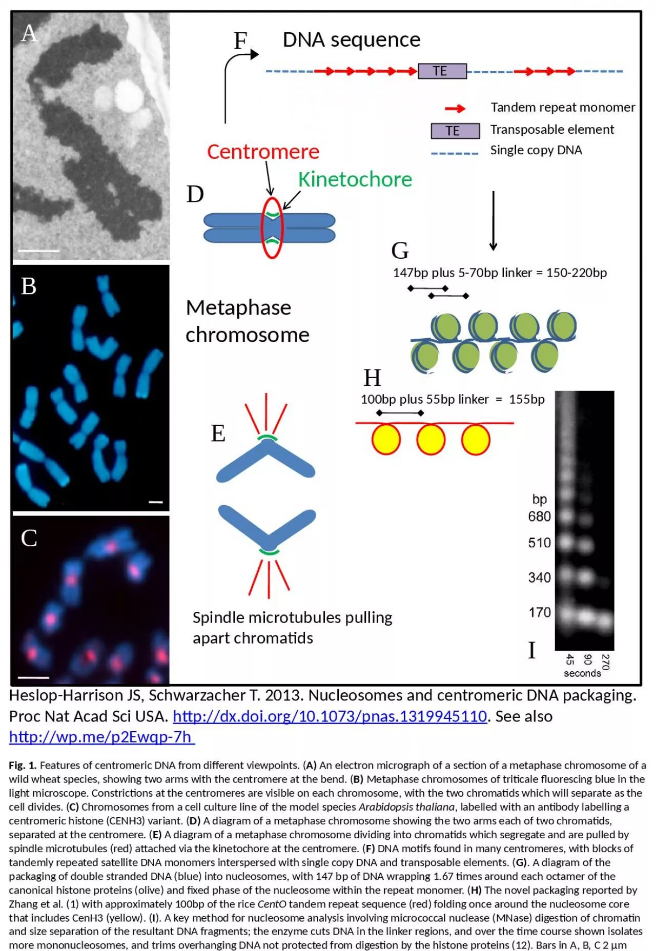
TE Tandem repeat monomer TE Transposable element Single copy DNA Spindle microtubules pulling apart chromatids Metaphase chromosome 147bp plus 570bp linker 150220bp 100bp plus 55bp linker 155bp ID: 1042487

chromosome dna centromere metaphase dna chromosome metaphase centromere repeat histone diagram chromatids centromeric nucleosome packaging linker species showing arms

Share:

Link:

Embed:

Download Presentation from below link

Download Presentation The PPT/PDF document "A B C Centromere DNA sequence" is the property of its rightful owner. Permission is granted to download and print the materials on this web site for personal, non-commercial use only, and to display it on your personal computer provided you do not modify the materials and that you retain all copyright notices contained in the materials. By downloading content from our website, you accept the terms of this agreement.


Presentation Transcript

1. ABCCentromereDNA sequenceTETandem repeat monomerTETransposable elementSingle copy DNASpindle microtubules pulling apart chromatidsMetaphase chromosome147bp plus 5-70bp linker = 150-220bp100bp plus 55bp linker = 155bpDEFGHIKinetochoreFig. 1. Features of centromeric DNA from different viewpoints. (A) An electron micrograph of a section of a metaphase chromosome of a wild wheat species, showing two arms with the centromere at the bend. (B) Metaphase chromosomes of triticale fluorescing blue in the light microscope. Constrictions at the centromeres are visible on each chromosome, with the two chromatids which will separate as the cell divides. (C) Chromosomes from a cell culture line of the model species Arabidopsis thaliana, labelled with an antibody labelling a centromeric histone (CENH3) variant. (D) A diagram of a metaphase chromosome showing the two arms each of two chromatids, separated at the centromere. (E) A diagram of a metaphase chromosome dividing into chromatids which segregate and are pulled by spindle microtubules (red) attached via the kinetochore at the centromere. (F) DNA motifs found in many centromeres, with blocks of tandemly repeated satellite DNA monomers interspersed with single copy DNA and transposable elements. (G). A diagram of the packaging of double stranded DNA (blue) into nucleosomes, with 147 bp of DNA wrapping 1.67 times around each octamer of the canonical histone proteins (olive) and fixed phase of the nucleosome within the repeat monomer. (H) The novel packaging reported by Zhang et al. (1) with approximately 100bp of the rice CentO tandem repeat sequence (red) folding once around the nucleosome core that includes CenH3 (yellow). (I). A key method for nucleosome analysis involving micrococcal nuclease (MNase) digestion of chromatin and size separation of the resultant DNA fragments; the enzyme cuts DNA in the linker regions, and over the time course shown isolates more mononucleosomes, and trims overhanging DNA not protected from digestion by the histone proteins (12). Bars in A, B, C 2 µmHeslop-Harrison JS, Schwarzacher T. 2013. Nucleosomes and centromeric DNA packaging. Proc Nat Acad Sci USA. http://dx.doi.org/10.1073/pnas.1319945110. See also http://wp.me/p2Ewqp-7h