/
Hydatid  liver disease : Hydatid  liver disease :

Hydatid liver disease : - PowerPoint Presentation

edolie
edolie . @edolie
Follow
27 views
Uploaded On 2024-02-03

Hydatid liver disease : - PPT Presentation

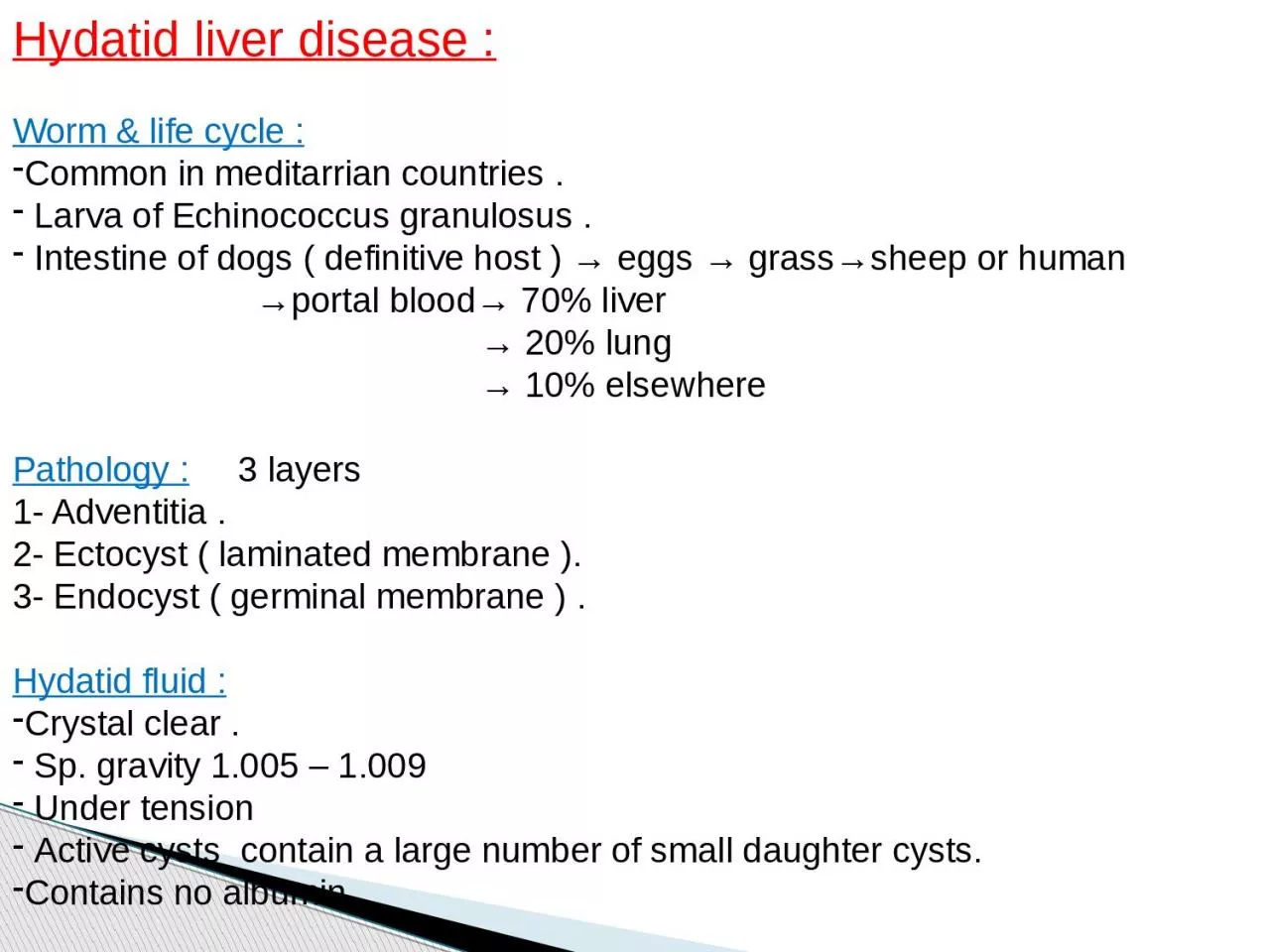
Worm amp life cycle Common in meditarrian countries Larva of Echinococcus granulosus Intestine of dogs definitive host eggs grasssheep or human portal blood 70 liver ID: 1044384

amp liver hepatic tumours liver amp tumours hepatic tissue jaundice mass malignant tumour clinical resection surgical features abdominal age

Share:

Link:

Embed:

Download Presentation from below link

Download Presentation The PPT/PDF document "Hydatid liver disease :" is the property of its rightful owner. Permission is granted to download and print the materials on this web site for personal, non-commercial use only, and to display it on your personal computer provided you do not modify the materials and that you retain all copyright notices contained in the materials. By downloading content from our website, you accept the terms of this agreement.


Presentation Transcript

1. Hydatid liver disease :Worm & life cycle :Common in meditarrian countries . Larva of Echinococcus granulosus . Intestine of dogs ( definitive host ) → eggs → grass→sheep or human →portal blood→ 70% liver → 20% lung → 10% elsewhere Pathology : 3 layers1- Adventitia .2- Ectocyst ( laminated membrane ).3- Endocyst ( germinal membrane ) . Hydatid fluid :Crystal clear . Sp. gravity 1.005 – 1.009 Under tension Active cysts contain a large number of small daughter cysts.Contains no albumin

2. Clinical features :1- symptomless 2- Abdominal mass .3- Pressure manifestation ; - Discomfort - Fullness & dyspepsia. - Jaundice4- Complications ; - Suppuration - Ruptured ….. daughter hydatids → intrabiliary → jaundice → acute cholangitis →Peritoneal → present as an acute abdomen after minor abdominal trauma. → pleural cavityCourse of the disease :1) Dead - calcified cyst .2) Enlarged gradually become manifested by its size and may float in the peritoneal cavity DDx mesenteric, pancreatic or renal cyst.3) Complication e.g. Rupture , infection .

3. Dx :1) US….multiloculated cyst 2) CT. scan & MRI…. floating membrane within the cysts3) Serological & immunological tests : a) Antibodies to hytadid antigen by ELISA . b) Casoni’s test 75% +ve . c) Compliment fixation test . Rx :Surgical Rx ; 1) open surgical Rx…A. Excision of the cysts B.Deroofing with evacuation of contents…. C. less likely liver resection. 2)Laparoscopic surgery might be beneficialScolicidal agents :1- Ethanol alcohol 95% . 2- Hypertonic saline 3- Silver nitrate 0.5% . 3- povidone iodine .Medical Rx ; 1) mebendazol 40 – 50 mg/kg. 2) Albendazol 10 – 15 mg/kg. 3) praziquantel 40 mg/kg

4. Complication of surgical treatment of hydatid cysts :Biliary leak → fistula Supuration → subhepatic , subphrenic or intrahepatic abscess Liver tissue damage and bleeding Recurrence Missed small deep cyst Injuries of adjacent organs Cholangitis Anaphylactic shock Contamination of peritoneal cavity. (careful, Albendazole& praziquantel) Wound infection

5. Liver tumours :.I - Benign tumour. II - Malignant tumour.I - Benign :A – Haemangioma : (vascular lesion) Most common . Often multiple of cavernous type . symptom less . If too large → mass – compressible .They have little if any malignant potential.Dx: -U/S …diagnostic… abnormal plexus of vessels. -CT… peripheral nodular enhancement on arterial phase then centripetal enhancement so it is delayed or slow contrast enhancement due to small vessel uptake. - P/c biopsy should be avoided → may bleed profusely Rx : If small & asymptomatic → No Rx If large and symptomatic → Rx is controversial (Embolisation , Lobe or segmental ressection or DXT to ↓ size) .Indication of Rx :Arterio venous shunting embarrass heart function .Misdiagnosis of malignant vascular tumour .

6. B – Hepatic adenoma : -premalignant-Women with contraceptive pills .-Develops in otherwise normal liver tissue.Dx: -U/S &CT → well circumscribed solid tumours. But, unfortunately difficult to differentiate from malignant tumours radiologically. So → -Angiography → well developed peripheral arterialisation of the tumoursRx . Lobe or segmental resection is Rx of choice. (premalignant)C – Focal nodular hyperplasia (FNH):-Focal overgrowth of functioning liver tissue supported by fibrous tissue stroma (hepatocytes & kupffer cells)- Middle age female with no association with underlying liver disease-Unusual & Unknown aetilogy.Dx -U/S → solid tumour, CT → central scarring & well-vascularised lesion (not specific) -Sulphur colloid liver scan →FNH +ve (kupffer cells) but adenoma & primary or metastatic tumours -ve (↓ ↓ kupffer cells )

7. II – Malignant liver tumours :Primary cancer :1- Hepatocellular carcinoma ( HCC ) : - one of the commonest tumour in the world specially UK - Association with chronic liver disease HBV & HCV → screening by U/S and AFP. -Middle age 3rd – 4th decades Male: female 8:1. - 80% of primary liver malignancy and 80% of HCC occur in cirrhotic liver. - May be multicentric . - ά Fetoprotein ↑ .Clinical features : -S&S of CLD: malaise and weakness , jaundice , ascites , portal hypertension. - S&S of Cancer : Mass ; wt. ↓ , anorexia ,,.Dx : CT. , US. , biopsy .Rx: 1. surgical resection (lobe or segment/s) 2. Liver transplantation (size, site and availability of donors)

8. 2- Cholangiocarcinoma : - Elderly age. – Patients with PSC - Fibrotic tumors → stricture often fibrous at the confluence of R&L hepatic ducts→ jaundice. - Distal b.d. CC → polypiod obstruct the lumen of b.d. Clinical features :-Painless obstructive jaundice-Enlarged tender liver , ↓ wt. , fever , astheniaDx -U/S → dilated intrahepatic but not extra-hepatic b.d.-Cholangiography → hilar stricture Brush cytology → tissue dx (2/3). -CT → ? Mass ( if infiltration liver parenchyma) - Angioraphy → (local spread to PV or HA).

9. RxSurgical resection + Chemo and radiotherapyPrognosis Very poor

10. Secondary liver tumours (Hepatic Deposits): Much more common-Multiple usually Sources :Intra abdominal : - GIT (colorectal), -Pancreas -Uterus & ovaries Extra abdominal: - Melanoma, Carcinoid tumours, Breast & Sarcoma .Dx:Dx of liver tumours as described before (U/S, CT….etc)Dx the primary- clinical exam, chest CT, bone scan, colonoscopy…. etc.Rx:1.Surgical resection2. If not resectable – systemic chemo (5 FU & folinic acid)Px:The 5-year survival rate after resection of solitary colorectal metas. is 35%

11. 2- Cholangiocarcinoma : - Elderly age. – Patients with PSC - Fibrotic tumors → stricture often fibrous at the confluence of R&L hepatic ducts→ jaundice. - Distal b.d. CC → polypiod obstruct the lumen of b.d. Clinical features :-Painless obstructive jaundice-Enlarged tender liver , ↓ wt. , fever , astheniaDx -U/S → dilated intrahepatic but not extra-hepatic b.d.-Cholangiography → hilar stricture Brush cytology → tissue dx (2/3). -CT → ? Mass ( if infiltration liver parenchyma) - Angioraphy → (local spread to PV or HA).