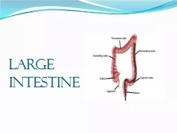
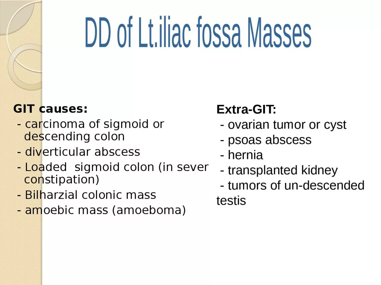
/
GIT causes:    - carcinoma of sigmoid or descending colon GIT causes:    - carcinoma of sigmoid or descending colon

GIT causes: - carcinoma of sigmoid or descending colon - PowerPoint Presentation

elina
elina . @elina
Follow
27 views
Uploaded On 2024-02-09

GIT causes: - carcinoma of sigmoid or descending colon - PPT Presentation

diverticular abscess Loaded sigmoid colon in sever constipation Bilharzial colonic mass amoebic mass amoeboma DD of Ltiliac fossa Masses ExtraGIT ovarian tumor or cyst ID: 1045464

diverticulitis diverticular colon bleeding diverticular diverticulitis bleeding colon sigmoid treatment abscess complicated resection patients bowel colonic attack high fistula

Share:

Link:

Embed:

Download Presentation from below link

Download Presentation The PPT/PDF document "GIT causes: - carcinoma of sigmoid or..." is the property of its rightful owner. Permission is granted to download and print the materials on this web site for personal, non-commercial use only, and to display it on your personal computer provided you do not modify the materials and that you retain all copyright notices contained in the materials. By downloading content from our website, you accept the terms of this agreement.


Presentation Transcript

1. GIT causes: - carcinoma of sigmoid or descending colon - diverticular abscess - Loaded sigmoid colon (in sever constipation) - Bilharzial colonic mass - amoebic mass (amoeboma) DD of Lt.iliac fossa MassesExtra-GIT: - ovarian tumor or cyst - psoas abscess - hernia - transplanted kidney - tumors of un-descended testis

2. Sigmoid colonLeft ovarySmall bowelRectum

3. Accounts for 14% of all cancer death (second to lung cancer)Risk factors include:Adenomatous polypsGenetic FactorDietary FactorsInflammatory Bowel DiseaseColon CancerEpidemiology and Risk factors

4. Colon Cancer abdominal pain & tenderness change in bowel habit blood in stool weight loss intestinal obstruction abd. & rectal exam. may reveal a mass .Signs & Symptoms

5. CBC, RFT, LFT. CT, MRIBiopsy, Histopathology.Sigmoidoscopy It is a must along with PR examination Can show any mucosal abnormality up to mid sigmoid colon (25 cm) Colonoscopy It visualize the entire colon.Colon CancerInvestigation

6. Always Remember ! Suspect Clinically . Confirm by Imaging . Prove by Histology .

7. It is surgical and require hemicolectomy Complications include:HemorrhageInjury of the bladder, ureter, small bowel, spleen, sexual function.Colon CancerManagement

8. COLONIC DIVERTICULAR DISEASE

9.

10.

11. * US Census Bureau, International Data Base, 2004 ( the extrapolations for Diverticular Disease are only estimates and may have limited relevance to the actual incidence of Diverticular Disease in any region)

12.

13. TYPES FALSE DIVERTICULA involves only protrusion of the mucosa through the muscularis propria of the colon most common TRUE DIVERTICULA a saclike herniation of the entire bowel wall

14.

15.

16. Protrusion occurs at the point where the NUTRIENT ARTERY or VASA RECTI penetrates through the muscularis propriaBreak in the integrity of the colonic wallCompression or erosionPERFORATION BLEEDINGPATHOPHYSIOLOGY

17. PATHOPHYSIOLOGY commonly affect the SIGMOID COLON due to:Relative high pressure zone within the muscular sigmoid colin.Higher amplitude contractions combined with constipated, high fat content stool within the sigmoid lumen results in the creation of these diverticula Related to retention of particulate material within the diverticular sac and formation of fecalith

18. Presentation, Evaluation, and Management

19. Diverticular BleedingHemorrhage from a colonic diverticulum is the most common cause of hematochezia in patients >60 years.Only 20% of patients with diverticulosis will have GI bleeding.Most bleeds are self-limited and stop spontaneously with bowel rest.Lifetime risk of rebleeding: 25%

20. Diverticular BleedingColonoscopy To localize the bleedingMay be both diagnostic and therapeutic in the management of mild to moderate diverticular bleedingAngiographyManagement of massive bleeding in a stable patientMesenteric angiography can localize the bleeding site and occlude the bleeding vessel successfully with a coil in 80% of the casesFollow up: Repetitive colonoscopy to look for evidence of colonic ischemiaSegmental resection of the colonTo eliminate risk of further bleedingIn patients on chronic blood thinners

21. Diverticular BleedingHighly selective coil embolizationSelective infusion of vasopressinTo stop hemorrhageComplications: MI, intestinal ischemiaRecurrence of bleeding in 50% of patients.

22. Diverticular BleedingSurgeryIndications: if patient is unstable or has had a 6-unit bleed within 24 hTotal abdominal colectomyPatients with presumed bleeding from diverticular disease requiring emergent surgery without localizationSurgical resection with primary anastomosisIn patients without severe comorbidities

23. Presentation, Evaluation, and Management

24. DIVERTICULITISUncomplicated – 75%Abdominal PainFeverLeukocytosisAnorexia/obstipationComplicated – 25%Abscess 16%Perforation 10%Stricture 5%Fistula 2%

25. DiverticulitisDiverticular perforationGeneralized peritonitis in <25% of cases(+) Abdominal distentionGiant diverticulum of the sigmoid(+) Air fluid level in the LLQ on plain abdominal film Mx: resection to avoid impending perforation

26. DiverticulitisDiagnosis is best made on CT.

27. DiverticulitisSuspected diverticulitis that does not meet CT criteria or is not associated with a leukocytosis or fever is not diverticular diseaseConditions that mimic diverticular disease:IBSOvarian cystEndometriosisAcute appendicitisPID

28. DiverticulitisBarium enema or colonoscopyShould be performed ~6 weeks after an attack of diverticular diseaseA sigmoid malignancy can masquerade as diverticular disease.Should not be performed in the acute settingHigher risk of colonic perforation associated with insufflation or insertion of barium-based contrast material under pressure.

29. DiverticulitisComplicated diverticular diseaseDiverticular disease associated with an abscess or perforation, and less commonly with a fistula.With fistula formationCommon locations include cutaneous, vaginal or vesicle fistulaePresent with either passage of stool through skin or vagina, or pneumaturiaColovaginal fistulae: more common in women who have undergone hysterectomy

30. Treatment of DiverticulitisComplicated diverticulitis = perforation,obstruction,abscess,fistula formation.□ Uncomplicated diverticulitis = Absence of the above complications

31. Treatment of Uncomplicated diverticulitisBowel rest or restrictionClear liquids or NPO for 2-3 daysThen advance dietBulk purgativesAntibioticsLifestyle modification : weight control, high fiber diet

32. Treatment Uncomplicated diverticulitis contd..Monitoring clinical coursePain should gradually improve several days (decrescendo)Normalization of temperatureTolerance of po intakeIF symptoms deteriorate or fail to improve with 3 days, then Surgery.After resolution of attack  high fiber diet with supplemental fiber is advised

33. Treatment Uncomplicated diverticulitis contd..Follow-up: Colonoscopy in 4-6 weeksFlexible sigmoidoscopy.PurposeExclude neoplasmEvaluate extent of the diverticulosis

34. Prognosis after resolution of uncomplicated diverticulitis□ 30-40% of patients will remain asymptomatic 30-40% of pts will have episodic abdominal cramps without frank diverticulitis20-30% of pts will have a second attack After a second attack  elective surgery

35. Prognosis after second attackSecond attackRisk of recurrent attacks is high (>50%)Some studies suggest a higher rate (60%) of complications (abscess, fistulas, etc) in a second attack and a higher mortality rate (2x compared to initial attack)Some argue elective surgery should be considered after a first attack inYoung patients under 40-50 years of age Immunosuppresed

36. Treatment of complicated diverticulitis

37. Treatment Complicated Diverticulitis: AbscessHINCHEY CLASSIFICATIONStage I Diverticulitis with associated pericolic abscessStage II Diverticulitis associated with distant abscess (retroperitoneal or pelvic)Stage III Diverticulitis associated with purulent peritonitisStage IV Diverticulitis associated with fecal peritonitis

38. STAGE i and STAGE ii is suitably managed with drainage and antibioticsSTAGE iii and STAGE iv usually requiring surgery

39. Treatment Complicated Diverticulitis: AbscessSmall <5 cm abscesses may resolve with antibiotic therapyPatient with larger abscesses or those who falls to improve with antibiotics should undergo CT guided percutaneous drainageColonic resection is indicated for those who develop either recurrent diverticulitis or another abscess

40. CT- GUIDED DRAINAGE OF DIVERTICULAR ABSCESS:Patient with abscess larger than or equal to 4 cmcan be managed with CT guided abscess drainage followed by elective surgery after resolution.

41. Complicated Diverticulitis: FistulasOccurs in up to 80% of cases requiring surgeryMajor typesColovesical fistula65%Colovaginal25%Coloenteric, colouterine10%

42. COLOVESICAL, COLOUTERINE AND COLOVAGINAL FISTULAE

43. Complicated Diverticulitis: Fistulas – Symptoms-Passage of gas and stool from the affected organColovesical fistula:pneumaturia, dysuria, fecaluria50% of patients can have diarrhoea and passage of urine per rectum

44. Complicated Diverticulitis: FistulasDiagnosisCT: thickened bladder with associated colonic diverticuli adjacent and air in the bladderBE: direct visualization of fistula track only occurs in 20-26% of casesFlexible sigmoidoscopy is low yield (0-3%)Some argue cystoscopy helpful

45. Complicated Diverticulitis: Treatment of colovesical FistulasTwo Approach to treat colovesical fistulaConservative: Without bowel resection by closing the fistula and interposing omentum between bowel and bladder.Conventional: Pinching off the affected bowel from the bladder, resect the sigmoid and perform end to end anastomosis.Bladder hole is left open and put urethral catheter for free drainage

46. Treatment of Complicated Diverticulitis:With generalised peritonitisSurgery is principally directed to control sepsis in the peritoneum and circulation.Vigorous resuscitation and antibiotic therapy.Opoid analgesia.Oxygen therapy.Urinary catheter to assess hourly urine out putResection of sigmoid colon and colorectal anastomosisPrimary resection and anastomosis after on table lavage in selected case.Hartman’s procedure : Resection of sigmoid colon with formation of end colostomy when condition do not favour primary anastomosis

47. Treatment of Complicated Diverticulitis:With obstruction/strictureSymptoms: pain,increasing constipation, passage of ribbon like stoolsHowever majority of patient presents with classic symptoms of large bowel obstructionDiagnosis is confirmed by ; patient’s history, physical examinations and radiological confirmation either by contrast enema or CT with oral/rectal contrast

48. Treatment of Complicated Diverticulitis:With obstruction/strictureConservative approach: Metallic stents to releive colonic obstruction.Endoluminal wall stents: shown to be safe and effective in decompressing obstructionSurgery: Hartman’s resection and resection with primary anastomosis rarely with loop colostomy is the procedure of choice.[Hartmann procedure is two stage procedure includes-Colostomy Sigmoid resection Rectal stump3 months later  colostomy takedown and colorectal anastomosis

49. Diverticular bleeding: SymptomsMost only have symptoms of bloating and diarrhea but no significant abdominal painPainless hematocheziaDiverticulitis rarely causes bleeding

50. Diverticular bleeding:Management□Most common cause of brisk hematochezia (30-50% of cases)15% of patients with diverticulosis bleed75% of diverticular bleeding stops without need for interventionPatients requiring less than 4 units : RBCs/ day  99% will stop bleedingRisk of rebleeding  14-38%

51. Diverticular bleeding: ManagementResuscitationLocalizationSupportive care with blood products

52. Diverticular bleeding: LocalizationRight colon is the source of diverticular bleeding in 50-90% of patientsPossible reasonsRight colon diverticuli have wider necks and domes exposing vasa recta over a great length of injuryThinner wall of the right colon

53. Diverticular bleeding:LocalizationColonoscopy after rapid preprationCan localize site of bleedingOffers possible therapeutic intervention (cautery, clip, etc)

54. Diverticular bleeding: Management ( Cauterization )

55. □ A site of active bleeding was identified□ Treated successfully with placement of two hemoclipsDiverticular bleeding:Management

56. Diverticular bleeding: SurgerySurgerySegmental resectionIf site can be localizedRebleeding rate of 0-14%Subtotal colectomyRebleeding rate is 0%High morbidity (37%)High mortality (11-33%)

57.