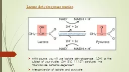
/
Figure 2 Figure 2. Modeling of the analysis of Plasmodium knowlesi lactate dehydrogenase Figure 2 Figure 2. Modeling of the analysis of Plasmodium knowlesi lactate dehydrogenase

Figure 2 Figure 2. Modeling of the analysis of Plasmodium knowlesi lactate dehydrogenase - PowerPoint Presentation

eliza
eliza . @eliza
Follow
66 views
Uploaded On 2023-05-21

Figure 2 Figure 2. Modeling of the analysis of Plasmodium knowlesi lactate dehydrogenase - PPT Presentation

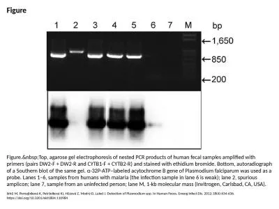
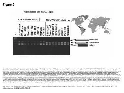
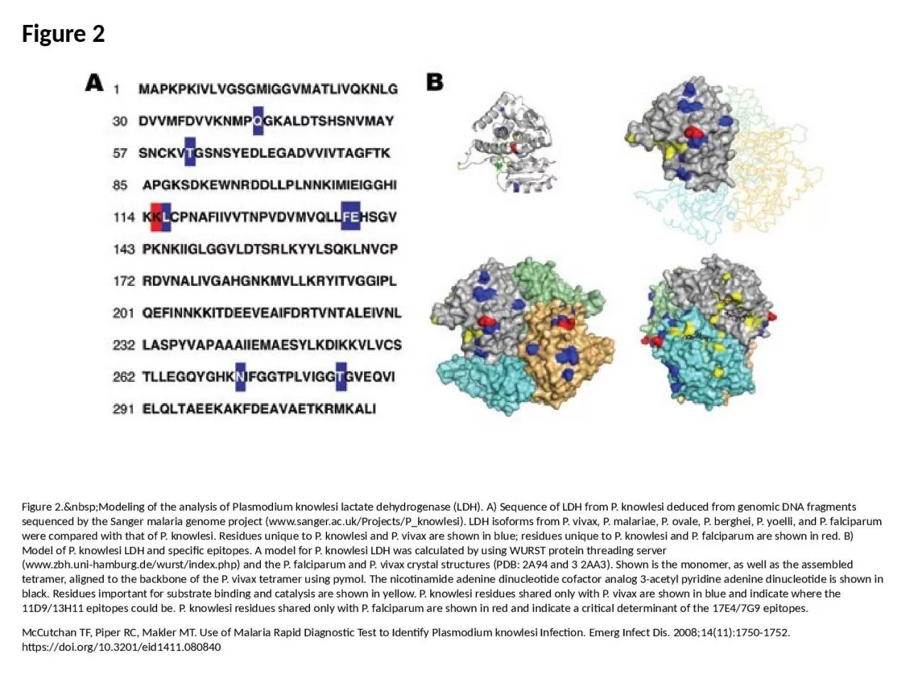
McCutchan TF Piper RC Makler MT Use of Malaria Rapid Diagnostic Test to Identify Plasmodium knowlesi Infection Emerg Infect Dis 2008141117501752 httpsdoiorg103201eid1411080840 ID: 998789

shown knowlesi vivax residues knowlesi shown residues vivax ldh falciparum epitopes plasmodium malaria unique tetramer red model wurst adenine

Share:

Link:

Embed:

Download Presentation from below link

Download Presentation The PPT/PDF document "Figure 2 Figure 2. Modeling of ..." is the property of its rightful owner. Permission is granted to download and print the materials on this web site for personal, non-commercial use only, and to display it on your personal computer provided you do not modify the materials and that you retain all copyright notices contained in the materials. By downloading content from our website, you accept the terms of this agreement.


Presentation Transcript

1. Figure 2Figure 2. Modeling of the analysis of Plasmodium knowlesi lactate dehydrogenase (LDH). A) Sequence of LDH from P. knowlesi deduced from genomic DNA fragments sequenced by the Sanger malaria genome project (www.sanger.ac.uk/Projects/P_knowlesi). LDH isoforms from P. vivax, P. malariae, P. ovale, P. berghei, P. yoelli, and P. falciparum were compared with that of P. knowlesi. Residues unique to P. knowlesi and P. vivax are shown in blue; residues unique to P. knowlesi and P. falciparum are shown in red. B) Model of P. knowlesi LDH and specific epitopes. A model for P. knowlesi LDH was calculated by using WURST protein threading server (www.zbh.uni-hamburg.de/wurst/index.php) and the P. falciparum and P. vivax crystal structures (PDB: 2A94 and 3 2AA3). Shown is the monomer, as well as the assembled tetramer, aligned to the backbone of the P. vivax tetramer using pymol. The nicotinamide adenine dinucleotide cofactor analog 3-acetyl pyridine adenine dinucleotide is shown in black. Residues important for substrate binding and catalysis are shown in yellow. P. knowlesi residues shared only with P. vivax are shown in blue and indicate where the 11D9/13H11 epitopes could be. P. knowlesi residues shared only with P. falciparum are shown in red and indicate a critical determinant of the 17E4/7G9 epitopes.McCutchan TF, Piper RC, Makler MT. Use of Malaria Rapid Diagnostic Test to Identify Plasmodium knowlesi Infection. Emerg Infect Dis. 2008;14(11):1750-1752. https://doi.org/10.3201/eid1411.080840