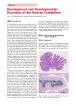
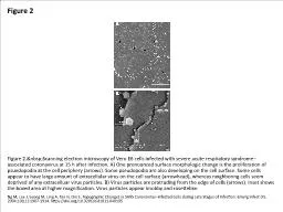
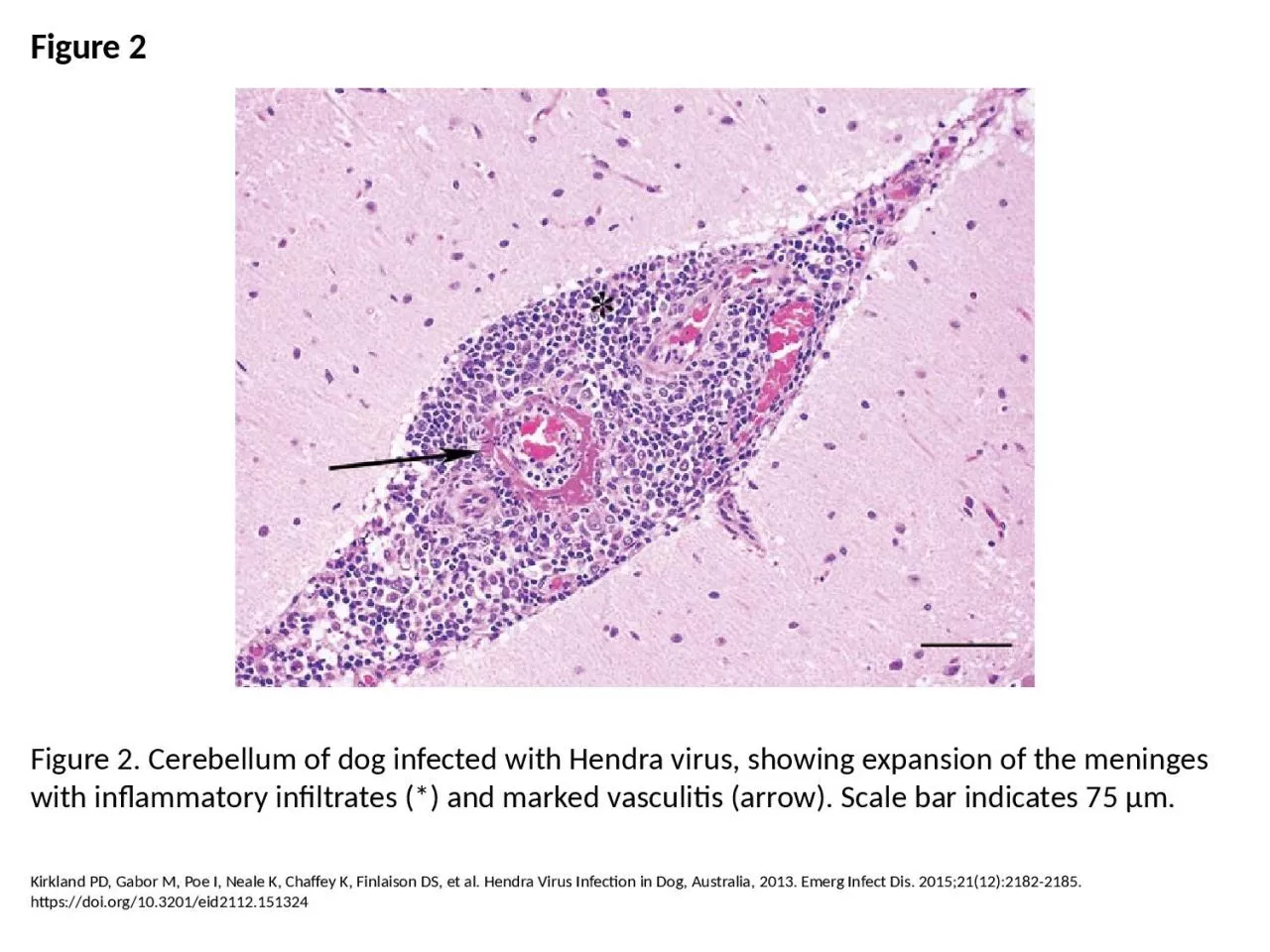
PPT-Figure 2 Figure 2. Cerebellum of dog infected with Hendra virus, showing expansion of

Author : erica | Published Date : 2023-05-22

Kirkland PD Gabor M Poe I Neale K Chaffey K Finlaison DS et al Hendra Virus Infection in Dog Australia 2013 Emerg Infect Dis 2015211221822185 httpsdoiorg103201eid2112151324

Presentation Embed Code

Download Presentation

Download Presentation The PPT/PDF document "Figure 2 Figure 2. Cerebellum of dog inf..." is the property of its rightful owner. Permission is granted to download and print the materials on this website for personal, non-commercial use only, and to display it on your personal computer provided you do not modify the materials and that you retain all copyright notices contained in the materials. By downloading content from our website, you accept the terms of this agreement.

Figure 2 Figure 2. Cerebellum of dog infected with Hendra virus, showing expansion of: Transcript


Kirkland PD Gabor M Poe I Neale K Chaffey K Finlaison DS et al Hendra Virus Infection in Dog Australia 2013 Emerg Infect Dis 2015211221822185 httpsdoiorg103201eid2112151324. Hope and Jen. Cerebellum. Brief anatomy and functions. Functions – DANISH. Pathology. Brief anatomy. What do the lobes of the cerebellum do?. Anterior cerebellum - unconscious proprioception. Posterior cerebellum - fine motor control. What is a Virus?. Viruses are microscopic particles (10 – 400 nm).. Viruses are made of genetic material (DNA or RNA) surrounded by a protein coating and sometimes an outer layer containing lipids.. Ágnes Nemeskéri. 2018. Semmelweis University. Department. of . Anatomy. , . Histology. . and . Embryology. nemeskeri.agnes. @. med.semmelweis-univ.hu. Function. of . cerebellum. C. erebellum is classically thought to control . 8.2.1SubdivisionTransverse fissures divide the cerebellum into lobes,lobules and folia,whereas paramedian sulci separatethe median vermis from the hemispheres (Fig.8.1).The flocculus and the nodule fo 1 CHAPTER 1 1 .1 The cerebellum which means ‘little brain’ is present in all vertebrate brains. Located underneath the posterior region of the skull and rostrocaudal to the occipital lobe, it /ISSN: 2160-4150/AJSC1205003 Review Article Meninges: from protective membrane to stem cell niche , Guido Fumagalli, Valeria Berton, Mauro Krampera, Francesco Bifari 93 n l y from the bite of mosquitoes (primarily the Culexspecies) infected with the virus. Mosquitoes become infected if they bite a bird thatcarries the virus. Infected mosquitoes then transmit West Nil – information for horse businesses Hendra virus is a sporadic infectious disease of horses caused by spill over of virus from flying foxes. Hendra virus can also spread to humans and other anim SKHMC. Location: . Posterior cranial . fossa. CEREBELLUM-LITTLE BRAIN. Connected to brain stem through 3 peduncles. Superior cerebellar peduncle-to midbrain. Middle cerebellar peduncle-to . pons. Inferior cerebellar peduncle-to medulla. Ng M, Lee J, Leong M, Ling A, Tan H, Ooi E. Topographic Changes in SARS Coronavirus–infected Cells during Late Stages of Infection. Emerg Infect Dis. 2004;10(11):1907-1914. https://doi.org/10.3201/eid1011.040195. Ng M, Lee J, Leong M, Ling A, Tan H, Ooi E. Topographic Changes in SARS Coronavirus–infected Cells during Late Stages of Infection. Emerg Infect Dis. 2004;10(11):1907-1914. https://doi.org/10.3201/eid1011.040195. Dr. Mohammad Rehan Asad. At the end of the lecture the student should be able to. Identify external features of cerebellum. Enumerate neurons, fibers, nuclei and layers of cerebellum. Identify connections of cerebellum. Cholecystography in a dog.. Bronchography in a sheep.. Myelography in a dog.. oesophagraphy after administration of 30 ml of Barium baste.. . GIT study showing presence of contrast material . ( Barium . lateral. . ventricles. Dr. Anna L. Kiss. Department. of . Anatómy. , . Histology. and . Embryology. Semmelweis University. Budapest. 2018. Prosencephalo. n. . (. yellow+red. ). telencephalon. .

Download Document

Here is the link to download the presentation.
"Figure 2 Figure 2. Cerebellum of dog infected with Hendra virus, showing expansion of"The content belongs to its owner. You may download and print it for personal use, without modification, and keep all copyright notices. By downloading, you agree to these terms.

Related Documents