/
Percivall   Pott 1714 – 1788 Percivall   Pott 1714 – 1788

Percivall Pott 1714 – 1788 - PowerPoint Presentation

fanny
fanny . @fanny
Follow
27 views
Uploaded On 2024-02-09

Percivall Pott 1714 – 1788 - PPT Presentation

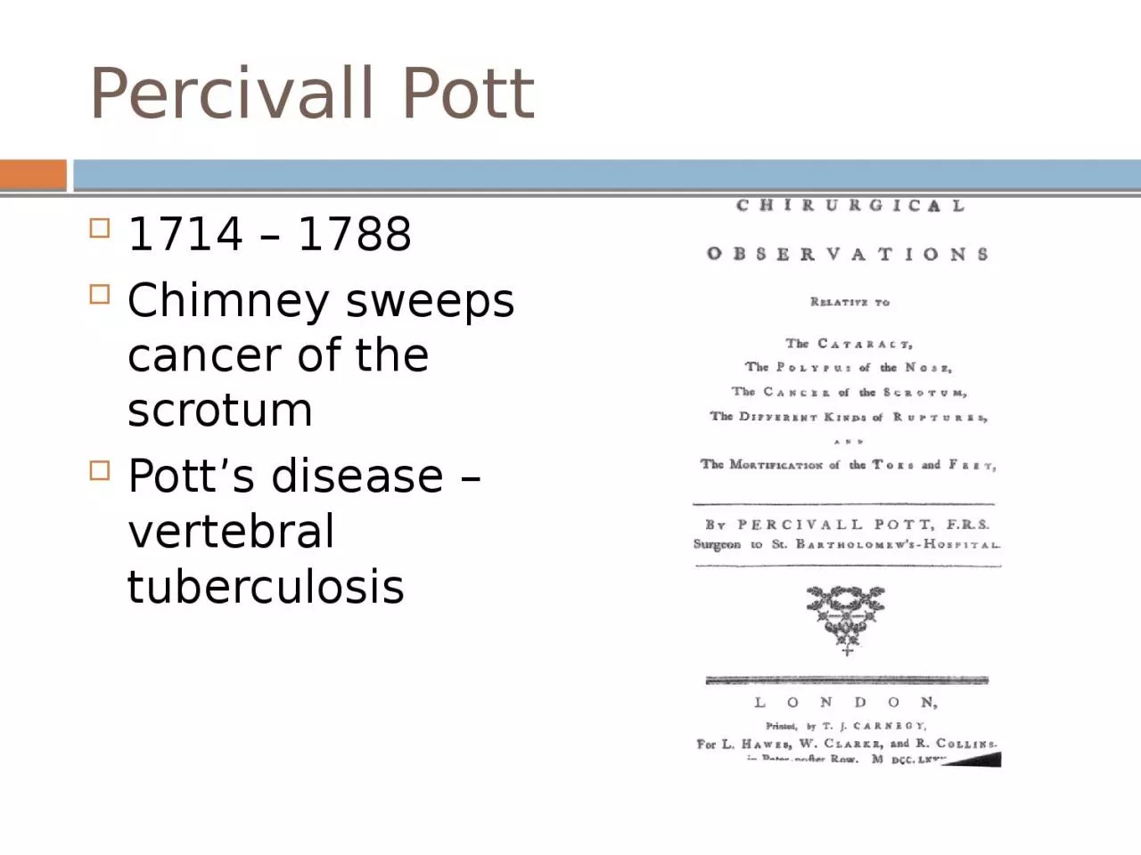
Chimney sweeps cancer of the scrotum Potts disease vertebral tuberculosis John Hunter 1728 1793 Papers at the Royal Society on experimental pathology including the use of a microscope ID: 1045079

cancer cell 1852 morbid cell cancer morbid 1852 1793 carcinoma diagnostic london cells pathology diagnosis royal atlas john vaginal

Share:

Link:

Embed:

Download Presentation from below link

Download Presentation The PPT/PDF document "Percivall Pott 1714 – 1788" is the property of its rightful owner. Permission is granted to download and print the materials on this web site for personal, non-commercial use only, and to display it on your personal computer provided you do not modify the materials and that you retain all copyright notices contained in the materials. By downloading content from our website, you accept the terms of this agreement.


Presentation Transcript

1. Percivall Pott1714 – 1788 Chimney sweeps cancer of the scrotum Pott’s disease – vertebral tuberculosis

2. John Hunter1728 – 1793 Papers at the Royal Society on experimental pathology, including the use of a microscopeDescribed inflammationHunterian Museum, Royal College of Surgeons, London

3. First Systematic Textbook of PathologyMatthew Baillie – 1761 – 1823 Nephew of John HunterThe Morbid Anatomy of Some of the Most Important Parts of the Human BodyMicroscopic pathology atlasPhysician of King George III

4. Thomas Hodgkin1798 – 1866 On Some Morbid Appearances of the Absorbent Glands and the Spleen“Lister’s compound microscope might lead to useful discoveries in the future.”

5. Joseph Recamier – 1774 – 1852 – metastasis Richard Bright – 1789 – 1858 – Kidney diseaseThomas Addison – 1793 – 1860 – Pernicious anemia

6. Cell TheoryRobert Hooke – 1635 – 1703 – cellMatthias Jacob Schleiden – 1804 – 1881 – botanist Theodor Schwann – 1810 – 1882 – zoologist

7. Cell TheoryJohannes Peter Müller1801 – 1858BerlinFather of medical microscopyMicroscopic criteria for benign and malignant tumorsÜber den Feinern Bau und die Formen der Krankhaften Geschwülste – On the Finer Structure and Form of Morbid Tumors

8. Cell TheoryRudolph Virchow1821 – 1902 The greatest figure in the history of PathologyDie Cellularpathologie“Omnis cellula e cellula” – all cells from cells

9. Herman Lebert – 1831 – 1878 Microscopic atlas

10. 1850sPathology developed as a separate specialtyMedical schools, Professors of PathologyMicroscope, diagnostic histopathology, neoplasiaFrance – laboratoriesGermany – universities

11.

12. MicroscopeFresh tissue, cut by hand, unstainedFormaldehyde fixation – Isaac Blum – 1833 – 1903 Paraffin embedding – Edwin Klebs – 1834 – 1913 Microtome – Minot – 1852 – 1914 Biological stainsHematoxalin – Franz BöhmerPaul Ehrlich – 1854 – 1915

13. AnaplasiaDavid Von Hansemann1858 – 1920

14. Grading, Carcinoma in SituAlbert Compton Broders1885 – 1964 Mayo Clinic

15. StagingCuthbert Esquire Dukes1890 – 1977 St. Mark’s Hospital, London

16. Pap SmearGeorge Papanicolaou – 1883 – 1962 January, 1928 – New Cancer Diagnosis – Betterment Conference Battle Creek Michigan1941 – The Diagnostic Value of Vaginal Smears in Carcinoma of the Uterus1943 – Diagnosis of Uterine Cancer by the Vaginal Smear1954 – Atlas of exfoliative Cytology