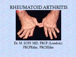
/
 RHEUMATOID ARTHRITIS Dr. M. SOFI MD; FRCP (London);  RHEUMATOID ARTHRITIS Dr. M. SOFI MD; FRCP (London);

RHEUMATOID ARTHRITIS Dr. M. SOFI MD; FRCP (London); - PowerPoint Presentation

faustina-dinatale
faustina-dinatale . @faustina-dinatale
Follow
347 views
Uploaded On 2020-04-04

RHEUMATOID ARTHRITIS Dr. M. SOFI MD; FRCP (London); - PPT Presentation

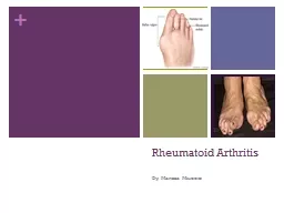
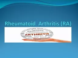
FRCPEdin FRCSEdin A chronic progressive disease causing inflammation in the joints and resulting in painful deformity and immobility especially in the fingers wrists feet and ankles ID: 775446

arthritis joint rheumatoid disease arthritis joint rheumatoid disease therapy joints patients early anti acpa patient swelling physical juvenile dmards

Share:

Link:

Embed:

Download Presentation from below link

Download Presentation The PPT/PDF document " RHEUMATOID ARTHRITIS Dr. M. SOFI MD; FR..." is the property of its rightful owner. Permission is granted to download and print the materials on this web site for personal, non-commercial use only, and to display it on your personal computer provided you do not modify the materials and that you retain all copyright notices contained in the materials. By downloading content from our website, you accept the terms of this agreement.


Presentation Transcript

Slide1

RHEUMATOID ARTHRITIS

Dr. M. SOFI MD; FRCP (London); FRCPEdin; FRCSEdin

Slide2

“A chronic progressive disease causing inflammation in the joints and resulting in painful deformity and immobility, especially in the fingers, wrists, feet, and ankles”.Pattern of joints affected — RA usually affects the same joints on both sides of the body. “In the early stages, rheumatoid arthritis typically affects small joints, especially the joints at the base of the fingers, the joints in the middle of the fingers, and the joints at the base of the toes. It may also begin in a single, large joint, such as the knee or shoulder, or it may come and go and move from one joint to another”.Joint symptoms — Usually begin gradually and include pain, stiffness, redness, warmth to the touch, and joint swelling.

RHEUMATOID ARTHRITIS

Slide3

Hands – The joints of the hands are often the very first joints affected by RA. Certain hand deformities can occur with longstanding RA. The fingers may develop swan neck and boutonniere deformities.Wrist – Most commonly affected joint. In the early stages of RA, it may become difficult to bend the wrist backward.Elbow – Swelling of this joint may compress nerves.

Swan neck deformity

Boutonniere deformity

Slide4

Shoulder – May be inflamed in the causing pain and limited motion.Foot – Often affected in the early. May be swollen and red.Ankle –RA of this joint may cause nerve damage, leading to numbness and tingling.Knee – RA may cause swelling of the knee, difficulty bending the knee, excessive looseness of the ligaments. May cause the Baker's cyst.Cervical spine – May cause a painful and stiff neck and a decreased ability to bend the neck and turn the head

Hallux

valgus and hammertoes

Sublaxation

metacarpophalyngeal

joints

Slide5

Pathogenesis not understood. An external trigger that triggers an autoimmune reaction, leading to synovial hypertrophy and chronic joint inflammation along with the potential for extra-articular manifestations.Synovial cell hyperplasia and endothelial cell activation are early events that progresses to uncontrolled inflammation and consequent cartilage and bone destruction.

Genetic factors and immune system abnormalities contribute to disease propagation.Abnormal production of cytokines, chemokines, and inflammatory mediators: TNF-aInterleukin -1, IL-6, IL-8,Transforming growth factor beta [TGF-ß]Fibroblast growth factor Platelet-derived growth factor (PDGF)

Pathophysiology: Rheumatoid arthritis

Slide6

The inflammation involved in rheumatoid arthritis can be intense. It is composed of mononuclear cells and can resemble a

pseudosarcoma

.

The hallmark of rheumatoid arthritis is a perivascular mononuclear cell infiltrate in the synovium. The early stages are noted to have plasma cells as well

Histologic

Findings

Slide7

Morning stiffness that lasts at least one hour and that has been present for at least six weeksSwelling of three or more joints for at least six weeksSwelling of the wrist, hand, or finger joints for at least six weeksSwelling of the same joints on both sides of the body

Changes in hand x-rays that are characteristic of rheumatoid arthritisRheumatoid nodules of the skinBlood test positive for rheumatoid factor and/or anti-citrullinated peptide/protein antibodies (ACPA)

DIAGNOSIS

 — There is no single test used to diagnose RA. Diagnosis is based upon many factors,

characteristic

signs and symptoms, the results of laboratory tests, and the results of x-rays.

Slide8

Slide9

Useful laboratory studies in suspected RA fall into 3 categories—Markers of inflammation,Hematologic parametersImmunologic parameter include following:Erythrocyte sedimentation rate (ESR)C-reactive protein (CRP) level

Complete blood count (CBC)Rheumatoid factor (RF) assayAntinuclear antibody (ANA) assayAnti−cyclic citrullinated peptide (anti-CCP)Anti−mutated citrullinated vimentin (anti-MCV) assays

Laboratory Studies

Slide10

Markers of inflammationThe ESR and the CRP level are associated with disease activity. The CRP value over time correlates with radiographic progression.Hematologic parametersCBC -- anemia of chronic disease correlates with disease activity; it improves with successful therapy. Hypochromic anemia suggests blood loss, commonly from GIT associated NSAIDs.

Thrombocytosis is common and is also associated with disease activity. Thrombocytopenia may be a rare adverse event of therapy and may occur in patients with Felty syndrome. Leukocytosis may occur but is usually mild. Leukopenia -- consequence of therapy or a component of Felty syndrome, which may then respond to DMARD therapy.

Laboratory

Studies

Slide11

Immunologic parametersRheumatoid factor — An antibody is present in the blood of 70 to 80 percent of people with RA. RF is not specific for RA but is also present in other connective tissue diseases, infections, and autoimmune disorders, as well as in 1-5% of healthy people. The presence of RF predicts radiographic progression of bone erosions, independent of disease activity.

RF values fluctuate with disease activity, though titers of RF generally remain high even in patients with drug-induced remissions.Anti-citrullinated peptide/protein antibody (ACPA) test — are more specific than RF for diagnosing RA. Anti-ACPA antibody tests may be positive very early in the course of disease. The test is positive in most patients with rheumatoid arthritis.

Laboratory

Studies

Slide12

Immunologic parametersANAs are present in about 40% of patients with RA.Assays for anti-citrullinated protein antibody (ACPA) are now used clinically for diagnosing RA.ACPA-positive and ACPA-negative RA may be 2 distinct disease subsets, with different underlying pathogeneses and risks for developing RA.

ACPA-positive patients may have a more erosive RA disease course than ACPA-negative patients.ACPA antibodies suggest a sensitivity and specificity equal to or better than those of RF.Presence of both anti-ACPA antibodies and RF is highly specific for RA. Presence of anti-ACPA antibodies, like that of RF, indicates a worse prognosis.

Laboratory

Studies

Slide13

For a definitive diagnosis of RA to rule out coexistent infection or crystal arthritis in an acutely swollen joint. In a new-onset monoarticular arthritis or an unusual flare up in a patient with RA may need joint aspiration and synovial fluid analysis.Analysis of synovial fluid includes Gram staining, cell count, culture, and assessment of overall appearance.

In RA, analysis typically reveals WBC count >2000/µL, generally in the range of 5000-50,000/µL. Usually, neutrophil predominance (60-80%) is observed (in contrast to mononuclear cell predominance in the synovium). Because of transport defect, glucose levels of synovial fluids (as well as pleural and pericardial fluids) in patients with RA are low compared with serum glucose levels.

Joint Aspiration indications

Slide14

Radiographic Feature

Peri-articular

osteopenia

Uniform symmetric joint space reduction

Marginal

subchondral

erosions

Joint

sublaxations

Joint destruction

Collapse

Ultrasound detects early soft tissue swelling

MRI has greatest sensitivity to detect

synovitis

and

marrow changes

Slide15

Soft-tissue swelling and early erosions in the proximal

interphalangeal

joints in a patient with rheumatoid arthritis of the hands.

Slide16

Widespread

osteopenia

, carpal crowding (due to cartilage loss), and several erosions affecting the carpal bones and metacarpal heads in particular in a child with advanced juvenile rheumatoid arthritis (also known as juvenile idiopathic arthritis).

Slide17

Subluxation

in the

metacarpophalangeal joints, with ulnar deviation,

Soft-tissue swelling and early erosions in the proximal

interphalangeal

joints 

Slide18

Soft-tissue swelling and early erosions in the proximal

interphalangeal joints in a patient with rheumatoid arthritis of the hands.

Well-defined bony erosions in the carpal bones and metacarpal bases in a patient with rheumatoid arthritis of the hands.

Slide19

T1-weighted sagittal

MRI

image of the cervical spine shows basilar invagination with cranial migration of an eroded odontoid peg.

Sagittal

T2- MRI of

cervical spine

Compromised

foramen

magnum

and there is increased signal intensity within upper cord; this is consistent with compressive

myelomalacia

.

Slide20

Extra-articular manifestations of Rheumatoid arthritis

Systemic

Musculoskeletal

Fever

Muscle wasting

Weight loss

Tenosynovitis

/Bursitis

Fatigue

Osteoporosis

Hematological

Ocular

Anaemia

Episcleritis

/

Scleritis

Thrombocytosis

Scleromalacia

Eosinophilia

Keratoconjunctivitis

sica

Vasculitis

Carditis

(30% in+ RA)

Digital

arteritis

Pericarditis/Myocarditis/

Ulcers

Conduction defects

Pyoderma

gangrenosum

Coronary vasculitis/

Granulomatous

aortitis

Slide21

FibromyalgiaLyme DiseaseMyelodysplastic SyndromeOsteoarthritisParaneoplastic SyndromesPolychondritis

Polymyalgia RheumaticaPsoriatic ArthritisSarcoidosisSjogren SyndromeSystemic Lupus Erythematosus (SLE)

Rheumatoid arthritis: Differential Diagnoses

Slide22

Directed toward the control of synovitis and the prevention of joint injury.The choice of therapies depends upon the Severity of disease activityWhen therapy is initiated Response of the patient to prior therapeutic interventions.Early recognition of diagnosis

Care by an expert in the treatment of RA Early use of disease-modifying antirheumatic drugs (DMARDs) for all patients diagnosed with RAImportance of tight control with target of remission or low disease activityUse of nonsteroidal anti-inflammatory (NSAIDs) and glucocorticoids, only as adjuncts to therapy

Treatment

:

Slide23

Focus on relieving pain Preventing damage/disability Patient education about thee disease Physical therapy for stretching and range of motion exercise

Occupational therapy for splints and adaptive devices Treatment should be started early and should be individualizedEarly aggressive treatment

Goals of management

RA

Slide24

Pretreatment evaluation —General testing for all patients include a baseline CBC, serum creatinine, aminotransferases, erythrocyte sedimentation rate (ESR), and C-reactive protein (CRP) in all patientsOphthalmologic screening for hydroxychloroquine use Testing for latent tuberculosis with skin testing or an interferon-gamma release assay prior to all biologic DMARDs

Treatment options RANSAIDSSteroidsDMARDsImmunosuppressive therapyBiological therapiesSurgery

Treatment for RA

Slide25

Briefly, these include:Patient educationPsychosocial interventionsRest, exercise, and physical and occupational therapyNutritional and dietary counseling

Interventions to reduce risks of cardiovascular disease, including smoking cessation, and of osteoporosisImmunizations to decrease risk of infectious complications of immunosuppressive therapies

NONPHARMACOLOGIC AND PREVENTIVE THERAPIES

Slide26

DMARDs:Nonbiologic DMARDsBiologic DMARDs, Orally-administered small molecule kinase inhibitorThe nonbiologic DMARDs HydroxychloroquineSulfasalazineMethotrexateLeflunomide

Biologic DMARDs, produced by recombinant DNA technology These medications are used include etanercept, infliximabcertolizumab which are all a class of drugs called TNF inhibitors. Other agents including anakinra, rituximab are often combined with methotrexate or other DMARADs to improve efficacy, generally target cytokines or their receptors

Choice of therapy

Slide27

In mildly active RA, initiate anti-inflammatory therapy with a NSAID for rapid symptomatic reliefBegin DMARD treatment with Hydroxychloroquine (HCQ) or Sulfasalazine (SSZ). In moderately to severely active RA, initiate anti-inflammatory therapy with either a NSAID or glucocorticoidBegin DMARD therapy with methotrexate .

In patients resistant to initial DMARD therapy (e.g., MTX), treat with a combination of DMARDs (eg, MTX plus either a TNF inhibitor or SSZ and HCQ)Switch the patient to a different DMARD of potentially comparable efficacy (eg, leflunomide or a TNF inhibitor), while also treating the active inflammation with ant-iinflammatory drug therapy.

Early use of DMARDs

Slide28

ASSESSMENT AND MONITORING

 — 

Patients should be seen on a regular basis for

clinical evaluation and monitoring of clinical and

laboratory assessment of disease activity and for

screening for drug toxicities.

Patient and clinician assessment of symptoms and

functional status

Evaluation of joint involvement and extra-

articular

manifestations

Laboratory markers

Imaging

Slide29

Juvenile Idiopathic Arthritis

Juvenile rheumatoid arthritis (JRA) is the most common chronic rheumatologic disease in children and is one of the most common chronic diseases of

childhood.

The

etiology is unknown, and the genetic component is complex, making clear distinctions between the various subtypes difficult

.

A

new nomenclature, juvenile idiopathic arthritis (JIA), is being increasingly used to provide better definition of subgroups.

Slide30

Signs and symptoms

History findings in children with JIA may include:Arthritis present for at least 6 weeks before diagnosis (mandatory for diagnosis of JIA)Either insidious or abrupt disease onset, often with morning stiffness or gelling phenomenon and arthralgia during the dayComplaints of joint pain or abnormal joint use

History of school absences or limited ability to participate in physical education classes

Spiking fevers occurring once or twice each day at about the same time of day

Evanescent rash on the trunk and extremities

Psoriasis or more subtle dermatologic

manifestations

Slide31

Physical findings

Physical finding :Arthritis: Defined either as intra-articular swelling on examination or as limitation of joint motion in association with pain, warmth, erythema of the joint; physical findings in JIA reflect the extent of joint involvementSynovitis: Characterized by synovial proliferation and increased joint volume; the joint is held in a position of maximum comfort, and range of motion often is limited only at the extremes

Slide32

Physical findings

Types of JIA include the following:Systemic-onset juvenile idiopathic arthritisOligoarticular juvenile idiopathic arthritisPolyarticular juvenile idiopathic arthritisPsoriatic arthritisEnthesitis-related arthritisUndifferentiated arthritis

Widespread osteopenia, carpal crowding (due to cartilage loss), and several erosions affecting the carpal bones and metacarpal heads in particular in a child with advanced juvenile rheumatoid arthritis

.

Slide33

Management

Management may include:Pharmacologic therapy with NSAIDs, disease-modifying antirheumatic drugs (DMARDs), biologic agents, or intra-articular and oral corticosteroidsPsychosocial interventionsMeasures to enhance school performance (e.g., academic counseling)Improved nutritionPhysical therapyOccupational therapy

American College of Rheumatology (ACR) criteria for complete remission are as

follows:

No inflammatory joint pain

No morning stiffness

No fatigue

No synovitis

No progression of damage, as determined in sequential radiographic examinations

No elevation of the ESR and CRP level

Slide34

Felty's syndrome is characterized by the combination of rheumatoid arthritis, splenomegaly and neutropenia. The condition is more common in those aged 50-70 years, and is more prevalent in females than males and more in Caucasians than blacks.Neutropenia – Neutropenia is present in all patients, with absolute neutrophil counts below 2000/microL.

Rheumatoid arthritis – The arthritis is typically severe, erosive, and seropositive for rheumatoid factor (RF) and/or anti-citrullinated peptide antibodies (ACPA) and is more frequently associated with extraarticular manifestationsSplenomegaly – Splenomegaly is present in most patients, although infrequently splenomegaly is undetectable in RA despite marked neutropenia

Felty's

syndrome

Slide35

Physical findings include:SplenomegalyHepatomegaly (mild)Lymphadenopathy Weight loss Rheumatoid nodules Sjögren syndrome Articular findings of long-standing RA – Joint deformities typical of RA, as well as synovitis (joint swelling and tenderness), which may be mild at presentation

Small-vessel inflammation (vasculitis) – Lower-extremity ulcers, palpable purpura and brownish pigmentary changes of the lower extremities, and periungual infarcts Signs of systemic vasculitis – Mononeuritis multiplex and extremity ischemia Other findings – Pleuritis, peripheral neuropathy, episcleritis, and signs of portal hypertension

Physical Examination

Slide36

Diagnostic ConsiderationsIn addition to the conditions listed in the differential diagnosis, other problems to be considered include the following: Chronic infection Drug reactions Other rheumatologic diseases Infiltrative diseases HIV infection Neutropenia with large granular lymphocytosis (LGL), also known as pseudo-Felty syndrome

Differential Diagnoses

Cirrhosis

Myeloproliferative Disease

Non-Hodgkin Lymphoma

Sarcoidosis

Sjogren

Syndrome

Systemic Lupus Erythematosus (SLE)

Tuberculosis

Slide37

WBC count and differential, which are crucial when determining the degree of granulocytopenia. Anemia and thrombocytopenia may result from hypersplenism. Mild elevations of ALP and transaminase levels may occur.Some 98% of patients with FS have high titers of RF. Antinuclear antibodies (ANAs), found in 67% of cases

Antineutrophil cytoplasmic antibodies (perinuclear pattern; p-ANCA), found in 77% of cases.ESR and serum immunoglobulin levels are invariably elevatedDiagnostic imagingRadionuclide studies, ultrasonography, or computed tomography (CT) may define the presence and extent of splenomegaly. The same modalities can also be used to assess patient response to therapy.

Laboratory and Imaging Studies

Slide38

ImmunosuppressantImmunosuppressive agents inhibit key factors in the immune system responsible for immune reactions.Methotrexate : It is very effective in treating rheumatoid arthritis (RA). Antirheumatic effects may take several weeks to become apparent. Methotrexate ameliorates symptoms of inflammation (e.g., pain, swelling, and stiffness). Cyclophosphamide is an antineoplastic alkylating agent and immunosuppressive agent. It reduces the numbers of B and T cells and increases the risk of infection.

Hematopoietic Growth FactorsCSFs stimulate production, maturation, and activation of neutrophils and increase migration and cytotoxicity of neutrophils.Most experience has been with the use of granulocyte CSF (G-CSF).Granulocyte-macrophage CSF (GM-CSF) stimulates division and maturation of earlier myeloid and macrophage precursor cells. It reportedly increases granulocytes in 48-91% of patients.Monoclonal Antibody: Rituximab Current data suggest that rituximab should be considered a second-line therapy in patients with refractory FS.

Treatment

Slide39

THANK

YOU

FOR

YOUR

ATTENTION