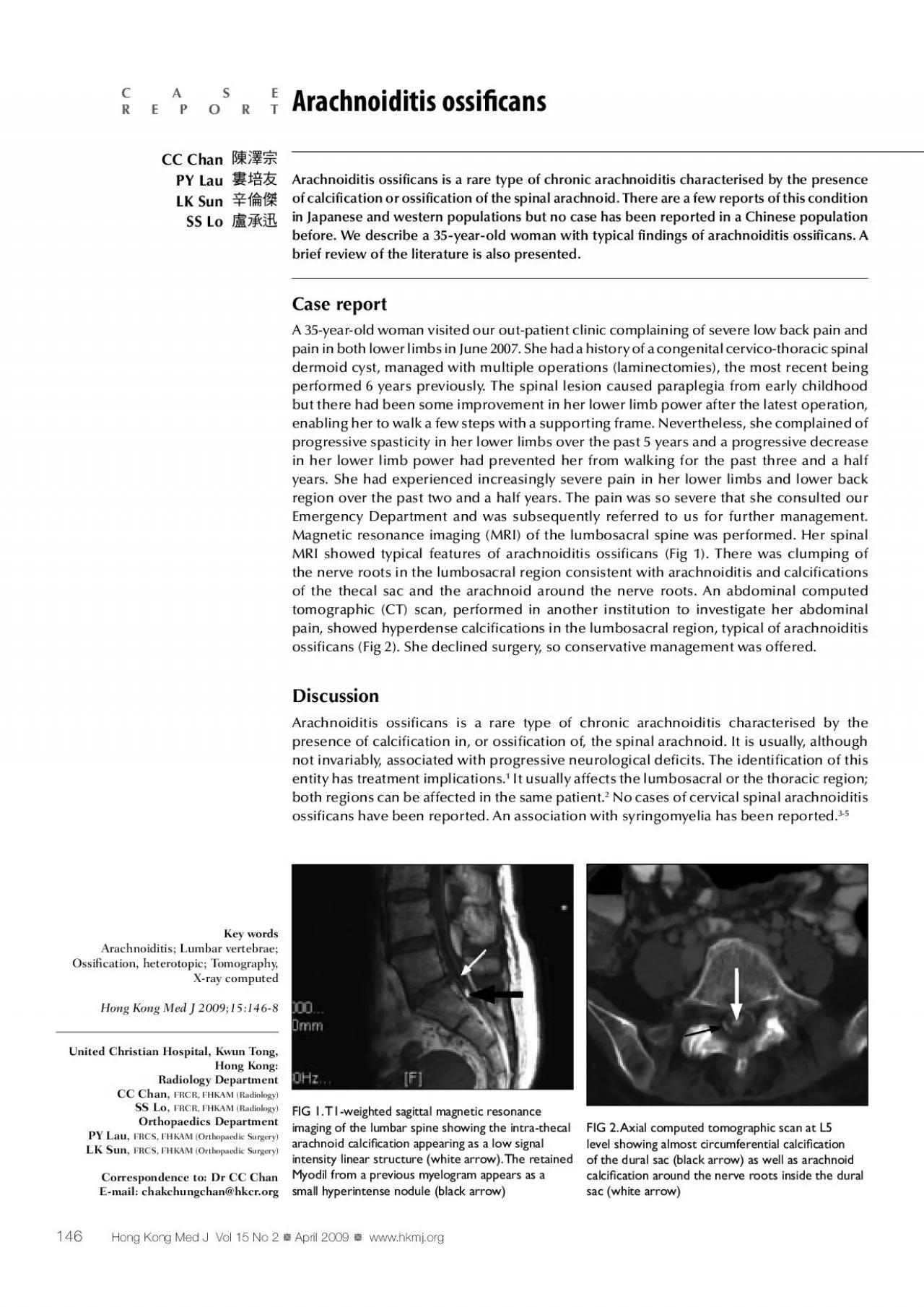
PDF-performed 6 years previously The spinal lesion caused paraplegia from

Author : gabriella | Published Date : 2022-08-16

but there had been some improvement in her lower limb power after the latest operation enabling her to walk a few steps with a supporting frame Nevertheless she

Presentation Embed Code

Download Presentation

Download Presentation The PPT/PDF document "performed 6 years previously The spinal ..." is the property of its rightful owner. Permission is granted to download and print the materials on this website for personal, non-commercial use only, and to display it on your personal computer provided you do not modify the materials and that you retain all copyright notices contained in the materials. By downloading content from our website, you accept the terms of this agreement.

performed 6 years previously The spinal lesion caused paraplegia from: Transcript


but there had been some improvement in her lower limb power after the latest operation enabling her to walk a few steps with a supporting frame Nevertheless she complained of progressive spastici. Fabien Koskas, Julien Gaudric. CHU Pitié-Salpêtrière, Paris, France. Ischémie . médullaire. Clampage médullaire. Hémodynamique. Hyperpression LCR. Hypoxie. PROTECTION MEDULLAIRE. Ischémie . médullaire. Abstract . Id. : IRIA- 1035. INTRODUCTION. Spinal Schwannomas and Meningiomas are the most common intradural extramedullary lesion and account for 45% of primary Spinal Neoplasms.. The clinical futures are usually Myelopathy and/or radiculopathy. Objectives. At the conclusion of this presentation the participant will be able . to:. Identify the components of the spine. Assess for spine and spinal cord injury. Discuss the initial management of the spinal cord injured patient. BJECTIVETo identify risk factors for Pseudomonas as the causative pathogen for hospitalized patients with cUTIStudy Design Retrospective cohort of hospitalized patients with cUTIadmitted to an academi BJECTIVETo identify risk factors for Pseudomonas as the causative pathogen for hospitalized patients with cUTIStudy DesignRetrospective cohort of hospitalized patients with cUTIadmitted to an academic James J. Lehman, DC, MBA, FACO. Associate Professor of Clinical Sciences. University of Bridgeport College of Chiropractic. Director. Community Health Clinical Education. University of Bridgeport. Learning Objectives. Introduction :. 2 General Classifications. Complete Lesion . A lesion to the spinal cord where there is no preserved motor or sensory function below the level of lesion. Incomplete Lesion. A lesion to the spinal cord with incomplete damage to the cord. There may be scattered motor function, sensory function or both below the level of lesion. ). Department of Neurology,. Zhongda. Hospital affiliated to Southeast . University. Nanjing. ,. China. Features of various part lesion. Motor cortex. Monoplegia: as motor cortex is spread over a large area, cortical lesion usually gives rise to monoplegia rather than hemiplegia. Spinal Cord. Location. Begins at the foramen magnum . Ends as conus medullaris at L. 1. vertebra. Functions. Provides two-way communication to and from the brain. Contains spinal reflex centers. Figure 12.30. June2009 www.hkmj.org 217 Case report We report a 38-year-old Chinese man (IV-2) with hereditary spastic paraplegia (HSP). He developed bilateral lower limb weakness in his late twenties and within Recibido: 2007; 18: 2007; 18: complication of spinal surgery, but the incidence of post-surgical SEHs that result in neurologic de�cits is extremely rare (0.1%). Patients that require mul 43 Chronic expanding organizing hematoma (CEH) occasionally mimics a soft tissue resection for musculoskeletal malignancy. Herein, we present a case of CEH which of the thigh, 19 months after surgery Riyesh T, Karuppusamy S, Bera BC, Barua S, Virmani N, Yadav S, et al. Laboratory-acquired Buffalopox Virus Infection, India. Emerg Infect Dis. 2014;20(2):324-326. https://doi.org/10.3201/eid2002.130358. Spinal cord is continuation of CNS contained within the bony spinal canal, from the foramen magnum at base of skull caudally to conus medullaris at level of L1 . . The three meningeal layers that surround the spinal cord continues below level of Ll as a fibrous tissue (filum teminale.

Download Document

Here is the link to download the presentation.
"performed 6 years previously The spinal lesion caused paraplegia from"The content belongs to its owner. You may download and print it for personal use, without modification, and keep all copyright notices. By downloading, you agree to these terms.

Related Documents