PDF-How to remove a corneal foreign body
Author : giovanna-bartolotta | Published Date : 2017-08-21
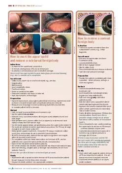
55 OC OBE 2005 HOW TO OPHTHALMIC PRACTICE Continued How to evert the upper eyelid and remove a subtarsal foreign body Indications To examine the upper tarsal conjunctiva
Presentation Embed Code
Download Presentation
Download Presentation The PPT/PDF document "How to remove a corneal foreign body" is the property of its rightful owner. Permission is granted to download and print the materials on this website for personal, non-commercial use only, and to display it on your personal computer provided you do not modify the materials and that you retain all copyright notices contained in the materials. By downloading content from our website, you accept the terms of this agreement.
How to remove a corneal foreign body: Transcript
Download Rules Of Document
"How to remove a corneal foreign body"The content belongs to its owner. You may download and print it for personal use, without modification, and keep all copyright notices. By downloading, you agree to these terms.
Related Documents