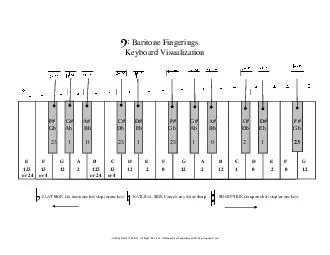
/
Silhouette Sign Silhouette Sign

Silhouette Sign - PowerPoint Presentation

giovanna-bartolotta
giovanna-bartolotta . @giovanna-bartolotta
Follow
397 views
Uploaded On 2016-06-08

Silhouette Sign - PPT Presentation

Frontal Xray Signs of Lobar Consolidation RUL loss of upper right mediastinal border RML loss of right heart border RLL loss of right hemidiaphram LUL loss of upper left ID: 353057

Share:

Link:

Embed:

Download Presentation from below link

Download Presentation The PPT/PDF document "Silhouette Sign" is the property of its rightful owner. Permission is granted to download and print the materials on this web site for personal, non-commercial use only, and to display it on your personal computer provided you do not modify the materials and that you retain all copyright notices contained in the materials. By downloading content from our website, you accept the terms of this agreement.


Presentation Transcript

Slide1

Silhouette SignSlide2

Frontal X-ray Signs of Lobar Consolidation

RUL

– loss of upper right

mediastinal

border

RML

– loss of right heart border

RLL

– loss of right

hemidiaphram

LUL

– loss of upper left

mediastinal

border

LINGULA

– loss of left heart border

LLL

– loss of left

hemidiaphramSlide3

Frontal X-ray Signs of Lobar Consolidation

SILHOUETTE SIGN

– for localizing lesion from frontal chest film. Intra-thoracic air space occupying lesions touching a soft tissue structure will obliterate the air soft tissue interface

Examples include:

Atelectasis

Pneumonia

NeoplasmSlide4

RLL Pneumonia

Density at the right lateral diaphragm

Obliteration of lateral diaphragm borderSlide5

RLL Pneumonia

Density at the mid diaphragm

Sharp margination at the major fissure (arrows)Slide6

Right Middle Lobe PneumoniaSlide7

LUL Lingular Pneumonia

Obliterated left cardiac borderSlide8

LUL

Lingular Pneumonia Lateral

Consolidation anterior to the major fissure

Compare to PA exam Slide9

LLL Pneumonia

Air space disease left lower lobe

Density behind heart

Obliteration of left diaphragm at edge of heart

Left heart border preservedSlide10

LLL Pneumonia

Note obliteration of the posterior portion of the left diaphragm (arrows)

Right diaphragm clearly seen