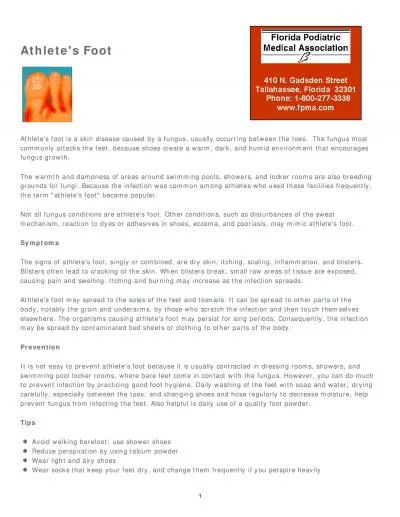
Site Name Scalp Tinea capitis Feet Tinea pedis Groin Tinea cruris Body Tinea corporis Nails Tinea unguium onychomycosis ID: 776549
Download Presentation The PPT/PDF document " Skin Conditions I Fungal Skin Infection..." is the property of its rightful owner. Permission is granted to download and print the materials on this web site for personal, non-commercial use only, and to display it on your personal computer provided you do not modify the materials and that you retain all copyright notices contained in the materials. By downloading content from our website, you accept the terms of this agreement.
Slide1
Skin Conditions I
Slide2Fungal Skin Infection/ Athlete’s foot
Site
Name
Scalp
Tinea
capitis
Feet
Tinea
pedis
Groin
Tinea
cruris
Body
Tinea
corporis
Nails
Tinea
unguium
(
onychomycosis
)
Slide3The fungus that causes the disease thrives in warm, moist conditions.The problem is more common in men than in women and responds well to OTC treatment.
Slide4Patient Assessment
Appearance
Severity
Location
Previous history
Medication
Slide5When to Refer
Severe, affecting other parts of the foot
Signs of bacterial infection
Unresponsive to appropriate treatment
Diabetic patients
Involvement of toenails
Slide6Management2 weeks
Pharmacists should instruct patients on how to use the treatment correctly to prevent recurrence.
Clean and dry your feet.
treatment must be continued
for
1–2 weeks after the disappearance of all signs of
infection to
ensure eradication of the fungus
Slide7Azoles (Miconazole, clotrimazole, ketconazole) TerbinafineGriseofulvin TolnaftateUndecenoates (e.g. zinc undecenoate)Hydrocortisone
Topical Anti-fungal
Slide8Eczema/dermatitis
Skin conditions
characterized by dryness, erythema, and itch of the skin, often with weeping and crusting
.
Contact dermatitis
(irritation or allergy).
Atopic eczema (with endogenous cause ).
Slide9Contact dermatitis & Atopic eczema
Typical eczema dermatitis
rash
Atopic eczema
.
Slide10Slide11Slide12Patient assessment
Age/distribution
Occupation/contact
History of hay fever/asthma
Severity…
Slide13Aggravating Factors
hay-fever season.
house dust or animal
danders
.
soaps or detergents and cold wind (dry the skin ).
Certain clothing such as
woollen
material can irritate the skin.
Cow’s milk, eggs and some food
colouring
.
Emotional factors, stress and
worry.
Antiseptic solutions applied directly to the skin or added to the
bathwater.
Slide14Medication
Contact dermatitis may be caused or made worse by
sensitisation
to topical medicaments.
Topically applied local
anaesthetics
, antihistamines, antibiotics and antiseptics can all provoke allergic dermatitis. Some preservatives may cause
sensitisation
.
Failed
medication need
referral
.
Slide15When to refer
Evidence of infection (weeping, crusting, spreading)
Severe condition: badly fissured/cracked skin, bleeding
Failed medication
No identifiable cause (unless previously diagnosed as eczema)
Duration of longer than 2 weeks
Slide16Treatment Timescale (1 week)
Non-pharmacological advices
Managing the
itch
Avoiding the
irritant
(e.g
.: wearing gloves to protect the skin
).
Maintaining the
skin integrity.
Slide17Pharmacological Treatment
Emollients
Topical corticosteroids
Antipruritics
Slide18Acne
Acne vulgaris is a common condition in young people.
It is not usually serious and resolves in most patients by the age of 25.
Peak incidence of acne is 14–17 years in females and 16–19 years in males.
The condition normally resolves in the majority of patients within 10 years of onset
.
Slide19Slide20Etiology
The hormonal changes
especially
the production of
androgens
.
Increased
keratin
and sebum production during
adolescence leads
to blockages of the follicles and the formation of
microcomedones
.
A
microcomedone
can develop into a
non-inflammatory
lesion (
comedone
), which may be open (blackhead) or closed (whitehead), or into an
inflammatory
lesion (papule, pustule or nodule).
Excess sebum encourages the growth of bacteria, particularly
Propionibacterium
acnes
, which are involved in the development of inflammatory lesions.
Slide21Patient Assessment
Age
Severity…
Affected areas
Medication
Slide22Severity
Mild
acne:
Patients with predominately
open and closed
comedones
with few inflammatory (
papulopustular
) lesions mainly confined to the face. Mild acne is therefore characterized by the presence of a few to
several
papules
and pustules the, but not
nodules
Moderate
acne:
A patient with moderate acne has
many inflammatory lesions
that are not confined to the face. Lesions are often painful and there is a possibility of mild
scaring.
Severe
acne:
A patient with severe acne has all the characteristics of moderate acne
plus the development of cysts
. Lesions are often widespread involving the upper back and chest. Scarring will usually result.
Slide23Slide24Slide25When to refer
Moderate and Severe acne
Failed medication
Suspected drug-induced
acne
Slide26Management
Benzoyl peroxide
(
2.5%, 5%, and 10% gels, lotion, cream)
Antibacterials
Topical
Retinoids
(
Adapalene
/
Differin
Gel 0.1%)
Slide27Practical points
Diet
Continuous treatment
Skin hygiene
Topical hydrocortisone and acne
Slide28Treatment timescaleimproved within 8 weeks
Only mild acne can be managed by the pharmacist using OTC products, moderate and severe acne should be referred.
Slide29Cold Sore
The virus responsible is the herpes simplex virus (HSV) of which there are two major types: HSV1 and HSV2.
Slide30Slide31Patient Assessment
Age and Duration
Symptoms and appearance
Location
Precipitating
factors
Previous history (help in
diagnosis
)
Medication
Slide32When to refer
Babies
and young children
Failure of an established sore to resolve
Severe or worsening sore (widespread)
History of frequent cold sores
Sore lasting longer than 2 weeks
Painless sore (like in oral cancer)
Patients with atopic eczema
Eye affected
Uncertain diagnosis
Immunocompromised
patient
Slide33Management
Aciclovir
and
Penciclovir
cream
Bland creams (e.g.
cetrimide
/
celavex
cream)
Hydrocolloid
patch
Slide34Advices to preventing cross-infection