/
THE ANATOMY OF THE DIGESTIVE SYSTEM THE ANATOMY OF THE DIGESTIVE SYSTEM

THE ANATOMY OF THE DIGESTIVE SYSTEM - PowerPoint Presentation

giovanna-bartolotta
giovanna-bartolotta . @giovanna-bartolotta
Follow
537 views
Uploaded On 2016-09-08

THE ANATOMY OF THE DIGESTIVE SYSTEM - PPT Presentation

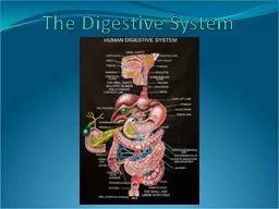
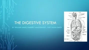
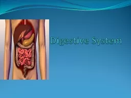
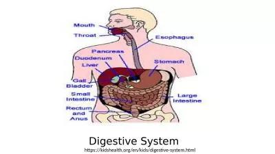
MOUTH Food enters here Tongue and teeth Lips cheeks palate Enzymes released in mouth to start digestion UVULA back of the mouth punching bag Small muscle that lifts to close the passage to the nasal cavities when we swallow ID: 462557

glands food swallow located food glands located swallow mouth digestive small stomach colon body secrete esophagus saliva villi digestion

Share:

Link:

Embed:

Download Presentation from below link

Download Presentation The PPT/PDF document "THE ANATOMY OF THE DIGESTIVE SYSTEM" is the property of its rightful owner. Permission is granted to download and print the materials on this web site for personal, non-commercial use only, and to display it on your personal computer provided you do not modify the materials and that you retain all copyright notices contained in the materials. By downloading content from our website, you accept the terms of this agreement.


Presentation Transcript

Slide1

THE ANATOMY OF THE DIGESTIVE SYSTEMSlide2

MOUTH

Food enters here

Tongue and teeth

Lips, cheeks, palate

Enzymes released in mouth to start digestion

UVULA

– back of the mouth (punching bag)

Small muscle that lifts to close the passage to the nasal cavities when we swallowSlide3

PHARYNX

Part of both digestive and respiratory systems

When we swallow, epiglottis lowers automatically and closes entrance to respiratory tract

Keeps food & liquid down and out of the lungs when you swallowSlide4

ESOPHAGUS

Connects pharynx to stomach and moves food

Located

behind

trachea

TRACHEA

– known as “windpipe”Slide5

STOMACH

J-shaped pocket

Small muscle, called

cardia

, located at junction of esophagus and stomach

Prevents food from travelling back up into the esophagus

Has a lining strong enough to hold up highly acidic environment needed to break down foodSlide6

SMALL INTESTINE

2 cm in diameter

3 m long

3 sections : duodenum, jejunum, ileum

Is folded several times to fit in our bodies

On the inside, it is lined with folds called

villiSlide7

LARGE INTESTINE

No villi here but it is made up of cells that secrete

mucus

MUCUS

– thick substance that lubricates the inner wall of the LI facilitating passage of solid residue

LI is made up of several parts : caecum, ascending colon, transverse colon, descending colon, rectumSlide8

DIGESTIVE GLANDS

Secrete substances that aid in the digestion of foodSlide9

SALIVARY GLANDS

3 pairs that produce saliva, moisten food and make it easier to swallow

Located at the upper and lower maxilla (jaws) and under the tongue

Each day, these glands secrete 1 to 1.5 L of saliva into the mouth!

Saliva plays a role in the digestion of carbs but also has

antibacterial

properties

ANTIBACTERIAL

– prevents growth of bacteriaSlide10

LIVER

Largest internal organ

Performs many tasks such as storing energy

Helps body get rid of toxins/poisons

Produces 1 L of bile each day

GALL BLADDER – located behind liver and is a small pouch that stores bile

Helps digest fats in the food you eatSlide11

PANCREAS

Looks like “corn on the cob”

Secretes over 1 L of pancreatic juices each day

Also secretes insulin which is a hormone that metabolizes sugar (regulates blood-glucose levels)

diabetesSlide12

GASTRIC GLANDS

Are scattered throughout the inner surface of stomach

Produces gastric juices in stomachSlide13

INTESTINAL GLANDS

Located at the bottom of the villi that line the inner surface of the S.I.

Produces even further chemical reaction on its way to L.I.Slide14

APPENDIX

Attached to cecum (L.I.)

Gets inflamed = appendicitis

May be removed

Could die if untreated due to ruptureSlide15

RECTUM

Last part of L.I.

Stores feces until it leaves the bodySlide16

ANUS

Exit point of the Digestive System

Feces leave the body