/
UNIVERSITY OF SOMALIA UNIVERSITY OF SOMALIA

UNIVERSITY OF SOMALIA - PowerPoint Presentation

giovanna-bartolotta
giovanna-bartolotta . @giovanna-bartolotta
Follow
397 views
Uploaded On 2017-11-05

UNIVERSITY OF SOMALIA - PPT Presentation

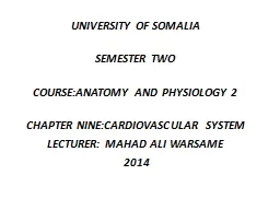
SEMESTER TWO COURSEANATOMY AND PHYSIOLOGY 2 CHAPTER NINECARDIOVASCULAR SYSTEM LECTURER MAHAD ALI WARSAME 2014 Key terms cardiovascular system a collection of organs that transport blood throughout the body consists of heart and blood vessels ID: 602833

heart blood vessels valves blood heart valves vessels valve left arteries pulmonary pressure ventricle cardiac ventricles layer rate beats

Share:

Link:

Embed:

Download Presentation from below link

Download Presentation The PPT/PDF document "UNIVERSITY OF SOMALIA" is the property of its rightful owner. Permission is granted to download and print the materials on this web site for personal, non-commercial use only, and to display it on your personal computer provided you do not modify the materials and that you retain all copyright notices contained in the materials. By downloading content from our website, you accept the terms of this agreement.


Presentation Transcript

Slide1

UNIVERSITY OF SOMALIA

SEMESTER TWO

COURSE:ANATOMY AND PHYSIOLOGY 2

CHAPTER NINE:CARDIOVASCULAR SYSTEM

LECTURER: MAHAD ALI WARSAME

2014Slide2

Key terms

cardiovascular system

:

a collection of organs that transport blood throughout the body (consists of heart and blood vessels)

heart

:

organ made of cardiac muscle with four chambers; responsible for pumping blood

atrium

:

chamber of the hear in the UPPER section

ventricle

: LOWER chambers of the heart

valves

: flap-like structures in heart near arteries the close to prevent blood from going backward

blood pressure

: the force with which blood is pumped through the arteriesSlide3

Key terms

Capillaries

:Smallest blood vessels; carries blood to the extremities

Veins

Medium sized blood vessels; carries blood to the heart

Arteries

Largest blood vessels; carries blood away from the heart

White blood cells

Cells

that fight disease

Red blood cells

Cells

that carry oxygenSlide4

Cardiovascular system

A closed system composed of the heart and blood vessels

The heart pumps blood

Blood vessels allow blood to circulate to all parts of the body

The general function of the cardiovascular system (

cvs

) is to deliver oxygen and nutrients and to remove carbon dioxide and other waste productsSlide5

The heart

is a hollow, muscular organ located between the lungs in the center and a bit to the left on the midline of the body its about 12 cm in length & about 9 cm in width.

Blood

: Blood is a viscous (thick) fluid that varies in color from bright to dark red, depending on how much oxygen it is carrying. its quantity differs with the size of the person; the average adult male, weighing 70 kg has about 5-6 liters of blood. Slide6

Con…

This volume accounts for about 8% of the total body weight. It is carried through a closed system of vessels pumped by the heart. The circulating blood is of fundamental importance in maintaining the internal environment in a constant state (homeostasis).Slide7

The heart is about the size of a clenched fist and comprises the following:

The heart composed of four layers:

Endocardium.

Myocardium.

Epicardium.

Pericardium.Slide8

endocardium

is the inner layer and is consists of endothelial tissue that lines the inner surface of the heart and the cardiac valves.

The

myocardium

is the middle layer and is composed of muscle fibers that enable the heart to pump.

Epicardium

is

the outer layer

,

is tightly adherent to the heart and the base of the great vessels.

A thin, fibrous, double-layered sac known as the

pericardium

surrounds the heart.Slide9

The

outer layer

is known as the

parietal pericardium

and the

inner layer

is called the

visceral pericardium

Between these two layers is a small amount of pericardial fluid (30 to 50 mL) that serves as a lubricant between the two layersSlide10
Slide11

The heart consists of four chambers:

a) right and left atrium

b) right and left ventricles.Slide12

Champers of heart

Four chambers of heart:

Atria

Receiving chambers

Right atrium

Left atrium

Ventricles

Discharging chambers

Right ventricle

Left ventricleSlide13

Heart valves

A heart valve normally allows blood to flow in only one direction through the heart. The four

heart

valve opens or closes incumbent on differential blood pressure on each side.Slide14

Con….

The four main valves in the heart are:

The two

atrioventricular

(AV) valves, the mitral valve (bicuspid valve), and the tricuspid valve, which are between the upper atria and the lower ventricles.

The two

semilunar

(SL) valves, the aortic valve and the pulmonary valve, which are in the arteries leaving the heart.Slide15

Cont..

Atrioventricular

valves

These are the

mitral

and

tricuspid valves

, which are situated between the atria and the ventricles and prevent backflow from the ventricles into the atria during systole.Slide16

Cont…

Semilunar

valves

Main articles:

Aortic valve

and

Pulmonary

valve

The

aortic and pulmonary valves are located at the base of the aorta and the pulmonary trunk respectively. These are also called the "

semilunar

valves". These two arteries receive blood from the ventricles and their

semilunar

valves permit blood to be forced into the arteries, and prevent backflow from the arteries into the ventricles. Slide17

Circulation of the blood

The blood passes through the tricuspid valve into the right ventricle, which then pumps the blood through the pulmonic valve into the pulmonary circulation.

After gas exchange in the lungs, oxygenated blood returns to the left atrium, passes through the mitral valve, enters the left ventricle, passes through the aortic valve, and finally enters the aorta.Slide18

circulatory Routes or Circuits

All the vessels together may be subdivided into two groups or

circuits: pulmonary and systemic.

1. Pulmonary circulation

: carry blood to and from the lungs.

They in include the pulmonary artery and its branches to the

capillaries in the lungs, as well as the veins that drain those

capillaries. The

pulmonary arteries

carry blood

low in

oxygen

from the right ventricle, while

the pulmonary veins

carry blood

high in oxygen

from the lungs into the left

atrium. This circuit functions to eliminate carbon dioxide

from the blood and replenish its supply of oxygen.

2. Systemic circulation

: it is the largest circulatory route. It

takes oxygenated blood from the left ventricle through the

aorta to all parts of the body.Slide19

Cardiac Output

Cardiac output

is the amount of blood pumped out of the ventricle .

The cardiac output in a resting adult is about 5 L per minute but varies greatly depending on the metabolic needs of the body. Slide20

Stroke

volume (SV) :

The amount of blood ejected by the left ventricle with

each heartbeat .

the heart rate is 60 to 80 beats per minute (

bpm

)

The average resting stroke volume is about 70 mL, and Cardiac output can be affected by changes in either stroke volume or heart rate.Slide21

Heart Rates

1.

Bradycardia

is a relatively slow heart rate of less than 60

beats/minute. During rest and sleep, the heart may beat

less than 60 beats/minute but usually does not fall below

50 beats/minute.

2.

Tachycardia

refers to a heart rate over 100

beats/minute.

3.

Sinus arrhythmia

is a regular variation in heart rate due

to changes in the rate and depth of breathing. It is normal

phenomenon. Slide22

4.

Premature beats

, also called extra systoles are beats that come in before the the expected normal beats. They may occur in normal persons initiated by caffeine, nicotine, or psycologic stresses. They are also common in persons with heart diseaseSlide23

Pulse

Pulse – pressure wave of blood

Monitored at “pressure points” where pulse is easily palpatedSlide24

Blood pressure

Measurements by health professionals are made on the pressure in large arteries

Systolic – pressure at the peak of ventricular contraction

Diastolic – pressure when ventricles relaxSlide25

Variations in blood pressure

Human normal range is variable

Normal

140–110 mm Hg systolic

80–75 mm Hg diastolic

Hypotension

Low systolic (below 110 mm HG)

Often associated with illness

Hypertension

High systolic (above 140 mm HG)

Can be dangerous if it is chronicSlide26

Patient Assessment:

Cardiovascular System

HEALTH HISTORY AND CLINICAL MANIFESTATIONS

For the patient experiencing an acute MI, the nurse obtains the health history using a few specific questions about the

onset and severity of chest discomfort, associated symptoms, current medications, and allergies.

At the same time, the nurse observes the patient’s general appearance and evaluates hemodynamic status (heart rate and rhythm, BP).

Slide27

Cardiac Signs and Symptoms

• Chest pain or discomfort (angina pectoris, MI,

valvular

heart disease) Shortness of breath or

dyspnea

(MI, left ventricular failure, HF)

• Edema and weight gain (right ventricular failure, HF)

• Palpitations (dysrhythmias resulting from myocardial ischemia, stress, electrolyte imbalance)Slide28

Fatigue (earliest symptom associated with several cardiovascular disorders)

• Dizziness and or loss of consciousness ( hypotension, dysrhythmias, ,cerebrovascular disorders)Slide29

Physical Exam

A) Inspection

General appearance

Jugular venous distension (JVD)

Skin

Extremities

B) Palpation

Pulses

C) Percussion

D) Auscultation

Good stethoscope

Positioning

Normal tones

Extra tones Slide30

HEART SOUNDS

The

normal heart sounds

, S1 and S2, are produced primarily by the closing of the heart valves.

The time between S1 and S2 corresponds to systole This is normally shorter than the time between S2 and S1 (diastole). As the heart rate increases diastole shortens.

S1—First Heart Sound.

Closure of the mitral and tricuspid valves creates the first heart sound (S1),

S2—Second Heart Sound. Closing of the aortic and pulmonic valves produces the second heart sound (S2).Slide31

Diagnostic Evaluation

Laboratory test(Cardiac Labs)

Chest X-ray

ECG

ECHOCARDIOGRAPHY(ECO)

Echocardiography is a noninvasive ultrasound test that is used to examine the size, shape, and motion of cardiac structures.Slide32

Blood vessels

Types of Blood Vessels

Arteries - vessels that transport blood away from the heart

Veins - return blood back to the heart

Capillaries - microscopic blood vessels that allow exchanges between blood and tissuesSlide33

Structure of a blood vessel

Tunica

externa

(adventitia) - outermost layer made of loose connective tissue. Serves to anchor, protect and prevent overstretching

Tunica media - middle layer composed of smooth muscle; functions in dilation and constriction of blood vessels

Tunica interna(

intima

) - innermost layer made of endothelium (s.squamous epithelium) Slide34

END