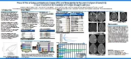
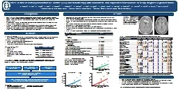
July 21 2021 Clinical presentation 70 yo male sp proximal and mid left SFA intervention on 07072021 with subsequent improvement in left lower extremity symptoms Now presents with R gt L lifestyle limiting claudication and rest pain Rutherford category 4 ID: 918911
Download Presentation The PPT/PDF document "Calcified Popliteal CTO ENDOVASCULAR LIV..." is the property of its rightful owner. Permission is granted to download and print the materials on this web site for personal, non-commercial use only, and to display it on your personal computer provided you do not modify the materials and that you retain all copyright notices contained in the materials. By downloading content from our website, you accept the terms of this agreement.
Slide1
Calcified Popliteal CTO
ENDOVASCULAR LIVE CASE
July 21, 2021
Slide2Clinical presentation
70
y.o
male s/p proximal and mid left SFA intervention on 07/07/2021 with subsequent improvement in left lower extremity symptoms.
Now presents with R > L lifestyle limiting claudication and rest pain (Rutherford category 4)
Exam shows
1+ dorsalis
pedis
pulse left side and
dopplerable
DP and PT on right side
Slide3PMH
CAD s/p CABG 3 V in 2013 and subsequent PCIs
Ischemic cardiomyopathy (EF 20%)
ESRD on HD
NIDDM
HTN HLD Ex smoker
Medications: Asa 81, Plavix, lipitor, Carvediolol, Glimeperide
Labs:
Hgb
/
Hct
10.1/31.5 INR 1.5
Plts
170
Slide4Peripheral angiogram
RLE Runoff
Slide5Peripheral angiogram
BTK DSA
Slide6Peripheral angiogram
Foot DSA
Indications Percutaneous intervention of FP-PCTO lesions is appropriate in patients with lifestyle-limiting claudication (Rutherford class [RC] 2-3) who have failed or are intolerant of pharmacological and exercise therapy.
Patients with CLI (RC 4-6).
Slide8StrategY
Access :
Antegrade
up and over
Antegrade
ipsilateral
Retrograde (Transpedal) Crossing: Ante grade wire .014” wire with .014” catheterEscalation to .018” or .035” wire to break the cap.
Revascularization:
Atherectomy
with PTA, DCB versus DES
Slide9Type 1: Antegrade or dual access, Type II : Dual access (sub intimal at distal cap)
Type III:
Antegrade
or Dual
access, Type IV : Retrograde
CTOP classification
CTOP: CTO crossing approach based on plaque cap morphology
Saab F, J
Endovasc
Ther
2018
Slide10PCTO Classification
Banerjee S, JOIC 2019
Slide11Antegrade techniques
STAR
(
Subinitmal
tracking and re entry): Continuing to advance a knuckled guidewire until it spontaneously re-enters the true lumen.
LAST
(Limited antegrade and subintimal tracking): Re-entry is achieved by using a guidewire with an acute(45°-60°, 1-2 mm) distal bend.
Antegrade
intraluminal wiring or ADR
Slide12Dissection is usually performed using a knuckle wire, whereas re-entry can be achieved by CART (Controlled Antegrade Retrograde Subinitmal
Tracking) or RCART techniques.
Inflating a balloon over the retrograde guidewire, followed by advancement of the
antegrade
guidewire into the distal true lumen is termed as CART.
RCART involves inflating a balloon over the
antegrade guidewire, followed by advancement of the retrograde guidewire into the proximal true lumen
Retrograde
C
ontrolled
A
ntegrade
R
etrograde Subintimal
T
racking
Slide13CART
Reverse CART
Double Balloon
Facilitated
Reentry
Retrograde
Techniques for Retrograde Crossing and Connecting
Slide14Tunneling:
involves passing of a retrograde or antegrade guidewire from an antegrade or retrograde catheter into a receiving catheter approaching from the opposite direction.
The re-back technique
refers to use of a needle-based re-entry device into a retrograde balloon to facilitate guidewire externalization.
Controlled dissection re-entry
is performed using specialized re-entry devices. The double-balloon technique involves inflation of two abutted balloon catheters advanced over the retrograde and antegrade guidewires. This may help merge the subintimal planes and facilitate true-lumen advancement of the antegrade or retrograde guidewires. SAFARI -
Subintimal arterial flossing with antegrade retrograde intervention
Advanced techniques
Slide15Banerjee S et al, JOIC 2019
Slide16Popliteal artery stenting (ETAP study)
Prospective, multi-center, randomized clinical trial.
246 pts were 1:1 randomized to nitinol stent (119) vs PTA (127).
Mean lesion length was 42.3 mm.
Primary patency rate was (64.2% vs 31.3%, p=0.0001) for BMS vs PTA @ 24 months.
TLR rate were (22.4% vs 59.5%, p=0.0001) for BMS @ 24 months.
Incidence of stent fracture was 4.6%
Significant improvement in ABI and Rutherford category was observed at 2 years in both groups.
Slide17Popliteal artery stenting (
Supera
Registry)
Retrospective, registry data review at a single center.
101 pts with 125 stents at the popliteal artery were followed for 12 months with US, XR, RC and ABI.
Primary Patency was 94.6 ± 2.3% and 87.7 ± 3.7% @ 6 and 12 months, respectively.
Secondary patency rates were 97.9 ± 1.5% and 96.5 ± 2.0% @ 6 and 12 months, respectively.
Mean ABI increased from 0.58 ± 0.15 to 0.97 ± 0.18, p<0.001)
No incidence of stent fracture was observed @ 15.2 month follow up.
Slide18Evidence for DES in Infra-popliteal vessels
Spiliopolous
et al, WJC, 2019
Slide19Adults with CLI (Rutherford category ≥4) and
infrapopliteal
lesions were randomized to receive PTA‐BMS (PTA with optional bailout stent) or DESs with paclitaxel.
5 years follow up.
Clinical end points were major amputation (above ankle level),
infrapopliteal
surgical or endovascular reintervention
, and death.
In total, 74 limbs (73 patients) were treated with DESs and 66 limbs (64 patients) were treated with PTA‐BMS.
The estimated 5‐year major amputation rate was lower in the DES arm (19.3% versus 34.0% for PTA‐BMS;
P
=0.091).
The 5‐year rates of amputation‐ and event‐free survival (survival free from major amputation or reintervention) were significantly higher in the DES arm compared with PTA‐BMS (31.8% versus 20.4%,
P
=0.043; and 26.2% versus 15.3%,
P
=0.041, respectively). Survival rates were comparable.
Spreen
ML et al, JAHA, 2017
Slide20Everolimus
-eluting Absorb bioresorbable vascular scaffold (BVS)
Mean lesion length was 20.1±10.8 mm
Single center prospective study of 48 patients
Primary patency estimates at 12, 24, and 36 months were 92.2%, 90.3%, and 81.1%; freedom from
clinically driven (CD)
- TLR
estimates were 97.2%, 97.2%, and 87.3% at the same time points.
Conclusion : Midterm follow-up demonstrates excellent safety, patency, and freedom from CD-TLR rates using the Absorb bioresorbable vascular scaffold below the
knee.
Varcoe
R et al, Journal of Endovascular Therapy 2018
Slide21Provisional with side branch wiring
Slide22Double Kiss (DK Crush technique), 6 Fr system
Kini
A, Practice Manual of Interventional Cardiology 2021
Slide23Mini crush technique
- 7fr system
Kini
A, Practice Manual of Interventional Cardiology 2021
Slide24THE CULOTTE TECHNIQUE
This is most suited to lesions where the SB and the distal MW are of similar caliber. The SB is stented first, followed by a POT and rewiring of the MV, aiming for a proximal cross of the stent struts. The stent struts are opened with a low profile balloon and the MV stented. A further POT is performed before rewiring of the SB to minimize the risk of
abluminal
wiring. KBI inflation is performed, sized to the distal vessels.
Slide25V stents
Slide26Slide27Slide28TAP technique (Bail out)
Kini
A, Practice Manual of Interventional Cardiology 2021
Slide29Thank You