/
Extracorporeal shock wave therapy in cubital tunnel Extracorporeal shock wave therapy in cubital tunnel

Extracorporeal shock wave therapy in cubital tunnel - PDF document

hadly
hadly . @hadly
Follow
343 views
Uploaded On 2022-08-26

Extracorporeal shock wave therapy in cubital tunnel - PPT Presentation

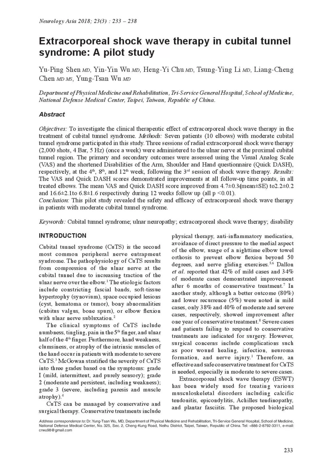
233 syndrome A pilot study YuPing Shen MD YinYin Wu MD HengYi Chu MD TsungYing Li MD LiangCheng Chen MD MS YungTsan Wu MD Department of Physical Medicine and Rehabilitation Tr ID: 941894

eswt x00660069 cuts tunnel x00660069 eswt tunnel cuts nerve patients study shock cubital syndrome ulnar therapy treatment dash wave

Share:

Link:

Embed:

Download Presentation from below link

Download Pdf The PPT/PDF document "Extracorporeal shock wave therapy in cub..." is the property of its rightful owner. Permission is granted to download and print the materials on this web site for personal, non-commercial use only, and to display it on your personal computer provided you do not modify the materials and that you retain all copyright notices contained in the materials. By downloading content from our website, you accept the terms of this agreement.


Presentation Transcript

233 Extracorporeal shock wave therapy in cubital tunnel syndrome: A pilot study Yu-Ping Shen MD , Yin-Yin Wu MD , Heng-Yi Chu MD , Tsung-Ying Li MD , Liang-Cheng Chen MD MS , Yung-Tsan Wu MD Department of Physical Medicine and Rehabilitation, Tri-Service General Hospital, School of Medicine, National Defense Medical Center, Taipei, Taiwan, Republic of China. Abstract To investigate the clinical therapeutic effect of extracorporeal shock wave therapy in the treatment of cubital tunnel syndrome. Methods: Seven patients (10 elbows) with moderate cubital tunnel syndrome participated in this study. Three sessions of radial extracorporeal shock wave therapy (2,000 shots, 4 Bar, 5 Hz) (once a week) were administered to the ulnar nerve at the proximal cubital tunnel region. The primary and secondary outcomes were assessed using the Visual Analog Scale (VAS) and the shortened Disabilities of the Arm, Shoulder and Hand questionnaire (Quick DASH), respectively, at the 4 th , 8 th , and 12 th week, following the 3 rd session of shock wave therapy. Results: treated elbows. The mean VAS and Quick DASH score improved from 4.7±0.3 ( mean ± SE) to2.2±0.2 and 16.6±2.1to 6.8±1.6 respectively during 12 weeks follow up (all p ) Conclusion: This pilot study revealed the safety and ef�cacy of extracorporeal shock wave therapy in patients with moderate cubital tunnel syndrome. Keywords: Cubital tunnel syndrome; ulnar neuropathy; extracorporeal shock wave therapy; disability Neurology Asia 2018; 23(3) : 233 – 238 Address correspondence to: National Defense Medical Center, No. 325, Sec. 2, Cheng-Kung Road, Neihu District, Taipei, Taiwan, Republic of China. Tel: +886-2-8792-3311, e-mail: crwu98@gmail.com INTRODUCTION Cubital tunnel syndrome (CuTS) is the second most common peripheral nerve entrapment syndrome. The pathophysiology of CuTS results from compression of the ulnar nerve at the cubital tunnel due to increasing traction of the ulnar nerve over the elbow. 1 The etiologic factors hypertrophy (synovium), space occupied lesions (cyst, hematoma or tumor), bony abnormalities (cubitus valgus, bone spurs), or elbow �exion with ulnar nerve subluxation. 2 The clinical symptoms of CuTS include numbness, tingling, pain in the 5 th �nger, and ulnar half of the 4 th �nger. Furthermore, hand weakness, clumsiness, or atrophy of the intrinsic muscles of the hand occur in patients with moderate to severe CuTS. 3 McGowan strati�ed the severity of CuTS into three grades based on the symptoms: grade I (mild, intermittent, and purely sensory); grade 2 (moderate and persistent, including weakness); grade 3 (severe, including paresis and muscle atrophy). 4 CuTS can be managed by conservative and surgical therapy. Conservative treatments include physical therapy, anti-in�ammatory medication, avoidance of direct pressure to the medial aspect of the elbow, usage of a nighttime elbow towel orthosis to prevent elbow �exion beyond 50 5,6 Dallon et al . reported that 42% of mild cases and 34% of moderate cases demonstrated improvement after 6 months of conservative treatment. 7 In another study, although a better outcome (80%) and lower recurrence (5%) were noted in mild cases, only 38% and 40% of moderate and severe cases, respectively, showed improvement after one year of conservative treatment. 8 Severe cases and patients failing to respond to conservative surgical concerns include complications such as poor wound healing, infection, neuroma formation, and nerve injury. 1 Therefore, an effective and safe conservative treatment for CuTS is needed, especially in moderate to severe cases. Extracorporeal shock wave therapy (ESWT) has been widely used for treating various musculoskeletal disorders including calcific tendonitis, epicondylitis, Achilles tendinopathy, and plantar fasciitis. The proposed biological 234 effects include neurogenesis, angiogenesis, anti-

in�ammation, production of growth factors, and the activation of progenitor cells and stem cells. 9 Axonal regeneration, proliferation of Schwann cells, and macrophage activation during myelin debris removal in nerve repair have also been described. 9 Recently, ESWT has been proven to be bene�cial in carpal tunnel syndrome (CTS). However, ESWT in the treatment of CuTS has not been assessed yet. Hence, we conducted a pilot study applying ESWT in patients with moderate CuTS. METHODS Participants We enrolled 7 patients (10 elbows), aged 35- 71 years, with a diagnosis of moderate CuTS. The diagnosis was based on clinical symptoms (numbness, tingling, pain in the 5 th �nger, and the ulnar half of the 4 th �nger) and electrophysiological studies. Patients’ symptoms and signs including motor weakness, had persisted for at least 6 months (McGowan grade 2). 4 The nerve conduction velocity �t the criter ia of a relative drop in conduction velocity more than 10 m/s of ulnar motor nerve across the elbow. 2 We excluded patients who had brachial plexus neuropathy, history of elbow trauma or operation, diabetes mellitus, malignancy, coagulopathy, pregnancy, active infection, or implanted pacemakers. All participants provided informed consent for this study. The trial was approved by the institutional review board of Tri-Service Genera l Hospital and registered at ClinicalTrials.gov (number Additional management during the study period was regulated in the patients. ESWT intervention Patients were seated and the target elbow was placed on the table with 40-50 degrees �exion, in a relaxed position. We identi�ed the ulnar nerve proximal to the inlet of the cubital tunnel where swelling of the nerve was observed with Tinel’s sign, using ultrasonography (MyLab™25Gold, Esaote, Genova, Italy) (Figure 1A). 11 Radial EWST (Pagani Elettronica, Milano, Italy) was administered with 2000 shots, 4 Bar pressure, and 5 Hz frequency by a single physician, using a probe perpendicular to the ulnar nerve (Figure 1B). 11,12 The participants received ESWT weekly for 3 consecutive weeks (total of 3 sessions). All patients tolerated the procedure well, without any analgesic agent. Outcome measurements The assessments were performed by a single physician, before treatment, and on the 4 th , 8 th , and 12 th week after the third ESWT session. Primary outcome The Visual Analog Scale (VAS) was used to quantify digital pain or paresthesia/dysthesia within one week. The scale ranged from 0 (painless) to 10 (severe pain). 14 Secondary outcome The Disabilities of the Arm, Shoulder and Hand questionnaire (DASH) has been widely used to evaluate upper extremity disorders, including CuTS. 15,16 We applied shortened DASH (Quick DASH), which exhibits similar effectiveness as DASH, for assessment. 17 The questionnaire contains an 11-item disability/symptom scale (each with 5 levels of response, ranging from 1 to 5). A higher score means more disability or more severe symptoms. Data analysis All the data were statistically analyzed by using the SPSS version 22.0 (SPSS Inc., Chicago, IL, Figure 1.(A) Detection of the swelling of the ulnar nerve (dotted circle) at cubital tunnel using ultrasonography. (B) The patient was seated and the target elbow was placed on the table with 40 to 50 degrees �exion. Radial shock wave therapy was applied with the probe perpendicular to the ulnar nerve. 235 Table 1: General data of study subjects Case no. Age (year) Sex Lesion side Duration (month) VAS Quick-DASH Baseline 4 th week 8 th week 12 th week Baseline 4 th week 8 th week 12 th week 1 Right 5 4 4 3 11.36 11.36 5.68 2 Male Left 4 3 3 2 11.36 5.68 5.68 4.55 3 Right 24 5 2 2 2 15.9 6.82 5.68 4.55 4 Left 24 4 2 2 2 11.36 2.27 4.55 4.55 5 Left 15 6 2 2 1 13.64 4.55 4.55 2.27 6 Right 6 5 2 2 2 13.64 2.27 2.27 7 Left 6 4 3 2 2 11.36 4.55 2.27 8 Male Left 6 6 5 4 4 2

2.73 22.73 13.64 11.36 9 Right 24 4 3 2 2 27.5 22.5 12.5 15 Left 24 4 3 2 2 27.5 22.5 12.5 15 Mean 47.9 27.9 4.7 2.9 2.5 2.2 16.64 11.66 7.50 6.75 VAS=Visual analog scale ; DASH=Disabilities of the Arm, Shoulder and Hand questionnaire. USA). The repeated-measures ANOVA followed by Bonferroni post hoc test and Wilcoxon singed- rank test were used for data at various follow-up. All statistical tests were two-tailed and a p-value less than 0.05 was considered as statistically RESULTS The mean age of the participants was 47.9 years. Three among them had bilateral CuTS (Table 1). Post-treatment, VAS decreased signi�cantly in all treated elbows at all follow-up time points (all p <0.01). The mean VAS score at baseline was ( mean ± SE), and improved to 2.9±0.3, 2.5±0.3, and 2.2±0.2, at the 4 th , 8 th , and 12 th week post-ESWT, respectively (Table 2 and Figure 2). The Quick DASH score also decreased in all cases with time; the mean point before management was 16.6±2.1 and decreased to 11.7±2.5, 7.5±1.3, and 6.8±1.6 at the 4 th , 8 th , and 12 th week post-treatment, respectively (Table 2 and Figure 2, all p < 0.01) . No patient exhibited complications or required additional management during the study. DISCUSSION To our knowledge, this is the �rst pilot study to assess the therapeutic effect of ESWT in patients with CuTS. The results showed significant improvement of symptoms and disability in all cases during the 12 weeks of follow-up. The mechanism of ESWT in promoting regeneration of peripheral nerves is unclear. Enhanced regeneration and functional recovery of injured nerves treated by ESWT have been observed in animal studies. 18,19 The biological mechanisms underlying these effects include activation of integrin-extracellular matrix coupling, rising proliferation of Schwann cells, enhanced macrophage activity, and molecular changes in neuron related expression of activating transcription factor 3 (ATF-3) and growth- associated phosphoprotein 43 (GAP-43). 9 Previous studies have also reported analgesic effects of ESWT, potentially contributing to symptom relief in peripheral neuropathy. ESWT can induce production of nitric oxide (NO) inhibiting NF- activation, and suppress inflammatory responses. NO can also directly impede pain conduction by opening potassium channels and reducing calcium in�ux into nerve cells, resulting in hyperpolarization. 10,22 The anti-in�ammatory action may also reduce swelling of the soft tissue surrounding the ulnar nerve and decrease the pressure in the cubital 236 tunnel. Moreover, ESWT may contribute to the reduction of calcitonin gene-related peptide (related to transmission of pain). 23 ESWT also inactivates the C �bers and increases activity within the descending inhibitory pathways from the brain stem that modulate gate-control of pain. Additionally, ESWT may promote angiogenesis by upregulating stromal-derived factor 1 (SDF- 1), vascular endothelial growth factor (VEGF), and its receptor Flt-1. These effects may improve tissue perfusion, thus, contributing to the clinical bene�ts 24,25 , as nerve ischemia caused by compression and stretching, may occur in CuTS. 3 In clinical practice, ESWT has been used for treating CTS. Seok et al . �rst reported signi�cant symptom relief and functional improvement at the 1 st and 3 rd month post-treatment with one session of ESWT. Consequently, other studies also con�rmed bene�cial effects at 3 to 6-months post-ESWT, for mild to moderate CTS, compared to nutraceutical treatment 13 , ultrasound 26 , or night orthosis. 27 Wu et al . also demonstrated the therapeutic and cumulative effects of ESWT has for mild-to-moderate CTS. 11,12 Although encouraging effects of ESWT in CTS have been reported, the therapeutic effects of ESWT in CuTS had not been examined in detail, prior to this pilot study. This is due to several dif

ferences between the pathophysiology of CTS and CuTS. First, in addition to direct compression of the ulnar nerve through the cubital tunnel, traction of the nerve and decrease of cubital tunnel space resulting from elbow �exion also play an important role in the development of CuTS. 3 However, the incidence of constant elbow �exion is less frequent and repetitive in patients with CuTS than CTS, in their daily activities. Hence, the spontaneous recovery and predictive Figure 2.Visual analog scale (VAS) and shortened Disabilities of the Arm, Shoulder and Hand questionnaire (Quick DASH) scores at baseline, and 4 th , 8 th , and 12 th week of follow-up. Compared to baseline, signi�cant improvements on both scales were noted at all follow-up timepoints (all p<0.01). ** p<0.01, ***p<0.001. Table 2: Outcome measures at baseline and 12-week follow-up Baseline ( mean ± SE) 4 weeks ( mean ± SE) 8 weeks ( mean ± SE) 12 weeks ( mean ± SE) VAS 4.7 ± 2.9 ± 2.5 ± 2.2 ± DASH 16.6 ± 2.1 11.7 ± 2.5** 7.5 ± 1.3*** 6.8 ± 1.6*** *Signi�cant difference compared to baseline (p<0.05), ** (p<0.01), ***(p<0.001). 237 outcome in CuTS are usually better than in CTS. Second, the perineural structures comprising the cubital tunnel such as Osborne’s ligament, medial collateral ligament, and medial epicondyle of the humerus are relatively hard, with less “cushions” like tendons in the carpal tunnel. Therefore, ESWT can reduce swelling of �exor tendons within the carpal tunnel contributing to greater decompression of the median nerve compared to that in CuTS. In addition to anatomical differences, different mechanisms of pathogenesis may contribute to the different therapeutic results observed due to ESWT in CuTS and CTS. The present pilot study has demonstrated similar therapeutic effects of ESWT in CuTS as CTS. Limited effects of conservative treatments, including orthoses, towel wrapping, patient education, and work modi�cation for moderate to severe CuTS have been reported. 3,7,8 We included patients with moderate severity and all of them showed improvement without complications, following 3 sessions of ESWT. The proportions are much more than the natural spontaneous recovery rates of about 50%. 3 No sham control was used in this pilot study. (The inclusion of a sham group was thought to be unacceptable to the patients before the therapeutic effect of ESWT for CuTS was con�rmed.) However, all patients were symptomatic for at least 6 months (mean 27.9 months), and spontaneous remission of symptoms could mostly be ignored in these chronic cases. Moreover, we can �nd the most signi�cant improvement in the �rst month after treatment. Hence, the effect of spontaneous recovery was considered to be small and the effect could be mainly attributable to ESWT. On the other hand, most patients in our study were newly diagnosed with CuTS. Hence, the potential for spontaneous remission still existed in these patients because they may have paid more attention to the pathophysiology of CuTS. Furthermore, placebo effect resulting from ESWT could also partially contribute to the therapeutic effect, especially in the initial days. Nevertheless, the placebo effect alone can hardly explain the signi�cant improvement lasting at least 12 weeks. Despite the above considerations, the therapeutic effects of ESWT for CuTS in our study may still be overestimated and a well-designed randomized controlled trial is recommended in the future. Some objective measurements, such as electrophysiological and ultrasonographic assessments, are good diagnostic tools, and may be used to assess the severity of CuTS. However, the relationship between these parameters and clinical outcomes in follow-ups is controversial. Indeed, Nathan et al . 28 and Gavin et al . 29 found that electrophysiological parameters are not consistentl

y correlated with clinical improvements. Duetzmann et al . reported that the cross-sectional area of the ulnar nerve did not correlate with the clinical outcomes after surgical decompression. The results of the study by Alblas et al . 31 also supported the aforementioned �ndings. Therefore, we did not apply these evaluations in our follow-up. Although it is valuable to measure muscle strength for post-treatment follow-up, we monitored the response using the Quick DASH score, which partially re�ect the muscle strength in daily activities. Izawa et al . 32 had noted that DASH scores correlated negatively with muscle strength. However, it would have been more bene�cial if we had tested muscle strength directly. This pilot study mainly focused on the clinical subjective outcomes and the feasibility of the therapy for a future trial. Further studies involving muscle strength, electrophysiological, and ultrasonographic evaluations are needed for a more comprehensive assessment. There are several limitations of this study. First, the number of cases in this pilot study is relatively small with only moderate CuTS. It does not represent the entire patient population. Further studies on patients with different grades of CuTS and larger sample size is suggested. Second, the follow-up duration is relatively short. Longer follow-up will be necessary to determine the long- term effects of ESWT. Signi�cant improvement of the VAS and quick DASH scores by week 12 continued to be observed. Therefore, we believe the bene�cial effects could persist beyond 12 weeks if longer follow-up periods were conducted. Third, spontaneous recovery and placebo effects of ESWT cannot be completely ruled out because there was no sham control group. Finally, it would be preferable to evaluate some objective parameters, such as electrophysiological and ultrasonographic features and muscle strength measurement, for a more comprehensive follow- up in the future study. In conclusion, this pilot study revealed that ESWT is a safe, non-invasive, and novel treatment to ease pain and improve disability in patients with moderate CuTS. Further well-designed studies are needed to validate these �ndings. DISCLOSURE Financial support: The study was supported by the Tri-Service General Hospital (grant no. TSGH-C106-089), Taipei, Taiwan, Republic of China. 238 Con�icts of interest: None REFERENCES Assmus H, Antoniadis G, Bischoff C, et al . Cubital tunnel syndrome–a review and management guidelines. Cent Eur Neurosurg 2011;72:90-8. Kroonen LT. Cubital tunnel syndrome. Orthop Clin North Am 3.Szabo RM, Kwak C. Natural history and conservative management of cubital tunnel syndrome. Hand Clin 2007;23:311-8. McGowan AJ. The results of transposition of the ulnar nerve for traumatic ulnar neuritis. J Bone Joint Surg Br 1950;32:293-301. 5.Boone S, Gelberman RH, Calfee RP. The management of cubital tunnel syndrome. J Hand Surg Am 6.Harder K, Diehm J, Fassola I. Current diagnostics and treatment of the cubital tunnel syndrome in Austria. GMS Interdiscip Plast Reconstr Surg DGPW 2016;5. Dellon AL, Hament W, Gittelshon A. Nonoperative management of cubital tunnel syndrome: an 8-year prospective study. Neurology 1993;43:1673-7. 8.Nakamichi K, Tachibana S, Ida M, Yamamoto S. Patient education for the treatment of ulnar neuropathy at the elbow. Arch Phys Med Rehabil Hausner T, Nógrádi A. The use of shock waves in peripheral nerve regeneration: new perspectives. Int Rev Neurobiol 2013;109:85-98. 10.Seok H, Kim SH. The effectiveness of extracorporeal shock wave therapy vs. local steroid injection for management of carpal tunnel syndrome: a randomized controlled trial. Am J Phys Med Rehabil 2013;92:327- 34. 11.Wu YT, Ke MJ, Chou YC, et al . Effect of radial shock wave therapy for carpal tunnel syndrome: A prospective randomized, double-blind, placebo- controlled trial. J Ortho

p Res 2016;34:977-84. 12.Ke MJ, Chen LC, Chou YC, et al . The dose- dependent ef�ciency of radial shock wave therapy for patients with carpal tunnel syndrome: a prospective, randomized, single-blind, placebo-controlled trial. Sci Rep 2016;6:38344. Notarnicola A, Maccagnano G, Tafuri S, Fiore A, Pesce V, Moretti B. Comparison of shock wave therapy and nutraceutical composed of Echinacea angustifolia, alpha lipoic acid, conjugated linoleic acid and quercetin (perinerv) in patients with carpal tunnel syndrome. Int J Immunopathol Pharmacol 14.Huskisson E. Measurement of pain. Lancet 1974;304:1127-31. 15.Hudak PL, Amadio PC, Bombardier C, et al . Development of an upper extremity outcome measure: the DASH (Disabilities of the Arm, Shoulder, and Hand). Am J Ind Med 1996;29:602-8. 16.Ido Y, Uchiyama S, Nakamura K, et al . Postoperative improvement in DASH score, clinical �ndings, and nerve conduction velocity in patients with cubital tunnel syndrome. Sci Rep 2016;6:27497. 17.Gummesson C, Ward MM, Atroshi I. The shortened disabilities of the arm, shoulder and hand questionnaire (Quick DASH): validity and reliability based on responses within the full-length DASH. BMC Musculoskelet Disord 2006;7:44. 18.Hausner T, Pajer K, Halat G, et al . Improved rate of peripheral nerve regeneration induced by extracorporeal shock wave treatment in the rat. Exp Neurol 2012;236:363-70. 19.Mense S, Hoheisel U. Shock wave treatment improves nerve regeneration in the rat. Muscle Nerve Mariotto S, de Prati AC, Cavalieri E, Amelio E, Marlinghaus E, Suzuki H. Extracorporeal shock wave therapy in in�ammatory diseases: molecular mechanism that triggers anti-in�ammatory action. Curr Med Chem 21.Ciampa AR, de Prati AC, Amelio E, et al . Nitric oxide mediates anti - in�ammatory action of extracorporeal shock waves. FEBS Lett 2005;579:6839-45. Hancock CM, Riegger-Krugh C. Modulation of pain in osteoarthritis: the role of nitric oxide. Clin J Pain Takahashi N, Wada Y, Ohtori S, Saisu T, Moriya H. Application of shock waves to rat skin decreases calcitonin gene-related peptide immunoreactivity in dorsal root ganglion neurons. Auton Neurosci Ito K, Fukumoto Y, Shimokawa H. Extracorporeal shock wave therapy as a new and non-invasive angiogenic strategy. Tohoku J Exp Med 2009;219:1-9. Hong JO, Park JS, Jeon DG, Yoon WH, Park JH. Extracorporeal Shock Wave Therapy Versus Trigger Point Injection in the Treatment of Myofascial Pain Syndrome in the Quadratus Lumborum. Ann Rehabil Med 2017;41:582-8. 26.Paoloni M, Tavernese E, Cacchio A, et al . Extracorporeal shock wave therapy and ultrasound therapy improve pain and function in patients with carpal tunnel syndrome. A randomized controlled trial. Eur J Phys Rehabil Med 2015;51:521-8. 27.Raissi GR, Ghazaei F, Forogh B, Madani SP, Daghaghzadeh A, Ahadi T. The Effectiveness of Radial Extracorporeal Shock Waves for Treatment of Carpal Tunnel Syndrome: A Randomized Clinical Trial. Ultrasound Med Biol 2017;43:453-60. Nathan P, Myers L, Keniston R, Meadows K. Simple decompression of the ulnar nerve: an alternative to anterior transposition. J Hand Surg Eur Vol Davis GA, Bulluss KJ. Submuscular transposition of the ulnar nerve: review of safety, ef�cacy and correlation with neurophysiological outcome. J Clin Neurosci 2005;12:524-8. Duetzmann S, Krishnan K, Staub F, Kang JS, Seifert V, Marquardt G. Cross-sectional area of the ulnar nerve after decompression at the cubital tunnel. J Hand Surg Eur Vol 2016;41:838-42. Alblas C, van Kasteel V, Jellema K. Injection with corticosteroids (ultrasound guided) in patients with an ulnar neuropathy at the elbow, feasibility study. Eur J Neurol 2012;19:1582-4. Izawa KP, Kasahara Y, Hiraki K, Hirano Y, Watanabe S. Relation between the Disability of the Arm, Shoulder and Hand Score and muscle strength in post-cardiac surgery patients. Diseases 2017;5:31. Neurology Asia September