/
Squamous cell carcinoma in situ of the cervix and placental  and Ins Squamous cell carcinoma in situ of the cervix and placental  and Ins

Squamous cell carcinoma in situ of the cervix and placental and Ins - PDF document

hailey
hailey . @hailey
Follow
342 views
Uploaded On 2022-08-21

Squamous cell carcinoma in situ of the cervix and placental and Ins - PPT Presentation

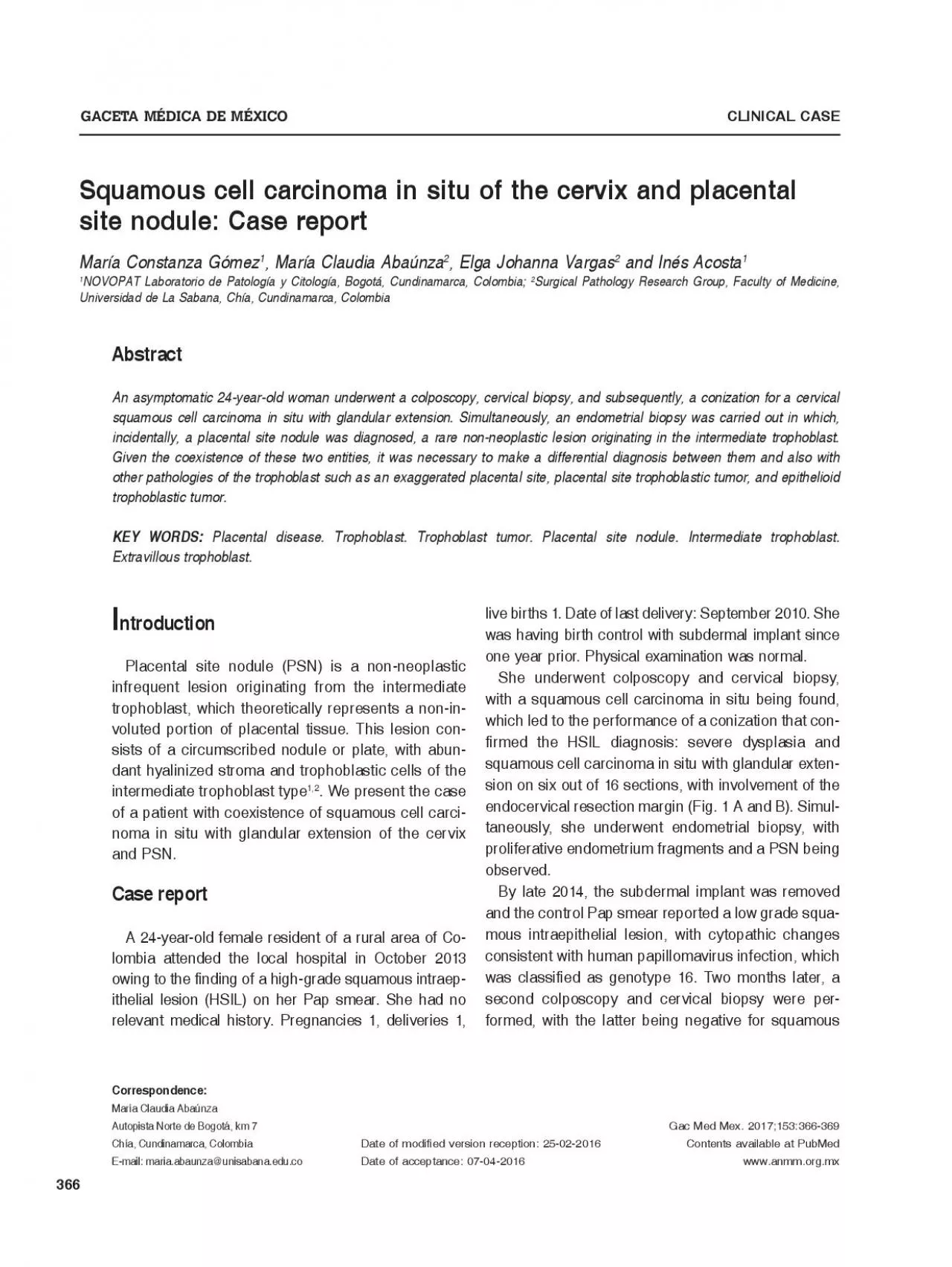
366 Placental site nodule PSN is a nonneoplastic infrequent lesion originating from the intermediate trophoblast which theoretically represents a noninvoluted portion of placental tissue This l ID: 939083

squamous placental site trophoblastic placental squamous trophoblastic site nodule cell carcinoma psn cervical positive trophoblast intermediate kurman cells situ

Share:

Link:

Embed:

Download Presentation from below link

Download Pdf The PPT/PDF document "Squamous cell carcinoma in situ of the c..." is the property of its rightful owner. Permission is granted to download and print the materials on this web site for personal, non-commercial use only, and to display it on your personal computer provided you do not modify the materials and that you retain all copyright notices contained in the materials. By downloading content from our website, you accept the terms of this agreement.


Presentation Transcript

366 Squamous cell carcinoma in situ of the cervix and placental and Inés AcostaSurgical Pathology Research Group, Faculty of Medicine, An asymptomatic 24-year-old woman underwent a colposcopy, cervical biopsy, and subsequently, a conization for a cervical Placental disease. Trophoblast. Trophoblast tumor. Placental site nodule. Intermediate trophoblast. GACETA MÉDICA DE MÉXICO Placental site nodule (PSN) is a non-neoplastic infrequent lesion originating from the intermediate trophoblast, which theoretically represents a non-in-voluted portion of placental tissue. This lesion con-sists of a circumscribed nodule or plate, with abun-dant hyalinized stroma and trophoblastic cells of the intermediate trophoblast type1,2. We present the case of a patient with coexistence of squamous cell carci-noma in situ with glandular extension of the cervix and PSN.A 24-year-old female resident of a rural area of Co-lombia attended the local hospital in October 2013 owing to the finding of a high-grade squamous intraep-ithelial lesion (HSIL) on her Pap smear. She had no relevant medical history. Pregnancies 1, deliveries 1, live births 1. Date of last delivery: September 2010. She was having birth control with subdermal implant since one year prior. Physical examination was normal.She underwent colposcopy and cervical biopsy, with a squamous cell carcinoma in situ being found, which led to the performance of a conization that con-firmed the HSIL diagnosis: severe dysplasia and squamous cell carcinoma in situ with glandular exten-sion on six out of 16 sections, with involvement of the endocervical resection margin (Fig. 1 A and B). Simul-taneously, she underwent endometrial biopsy, with proliferative endometrium fragments and a PSN being observed.By late 2014, the subdermal implant was removed and the control Pap smear reported a low grade squa-mous intraepithelial lesion, with cytopathic changes consistent with human papillomavirus infection, which was classified as genotype 16. Two months later, a secon

d colposcopy and cervical biopsy were per-formed, with the latter being negative for squamous M.C. Gómez, et al.: Cervical carcinoma and placental nodule intraepithelial lesion. The patient is currently breast-feeding her second child.PSN occurs in childbearing age women and, usually, it is an incidental finding of endocervical cu-rettages, cervical and endometrial biopsy and hyster-ectomy1,4,5. It is mainly located at the endometrium or the cervix, and rarely at the uterine tube. It is usually detected several months or years after a pregnancy, with an average of 3 yearsrhagic nodule is appreciated, with a diameter of 1 to 14 mm, although it rarely exceeds 4 mm; it can occur ma with intermediate trophoblast-type cells distributed in groups or cords, or individually displayed with absence or sparse atypical mitoses. The cells vary in size; the small ones are mono- or binucleated, with a clear, glycogen-rich cytoplasm, and the large ones have broad acidophilic or amphiphilic cytoplasms, with tial inflammatory infiltrate composed of lymphocytes (Fig. 2 B). and cyto-keratin 18, with focal or negative expression for hPL and CD146 (Mel-CAM). They are usually negative for ogy and immunohistochemical (IHC) characteristics, PSNs originate from the intermediate trophoblast of the chorionic type, and it is suggested that they are the Most important differential diagnoses include (Table 1). EPS is diagnosed after a normal pregnancy, an ectopic pregnancy or a PSN composed of intermediate type trophoblastic cells trophoblastic Figure 1. A: squamous cel carcinoma in situ of the cervix (hematoxylin & eosin, x10). B: glandular extension (hematoxylin & eosin, x20). Gaceta Médica de México. PSN trophoblastic cells are positive for P63 (IHC, x20) ( EPSPSNPSTTETTSCCHistogenesisImplantation site intermediate trophoblastintermediate trophoblastintermediate trophoblastintermediate trophoblastNon-trophoblasticGrowth patternNot arranged in Circumscribed circumscribed CellularityHighLowHighHighHighCytologi

cal atypiaGeneralized Focal/sparseGeneralizedGeneralizedGeneralizedMitotic figuresAbsentAbsent to rareCommonCommonCommonStromaFibrin depositAbundant Deposit of fibroid NecrosisAbsentOccasional, Chorionic villiPresent AbsentAbsentAbsentAbsentImmunohistochemistry CK 18Positive. DiffusePositive. DiffusePositive. DiffusePositive. DiffuseNegativeCD146(Mel-CAM)Positive. DiffuseNegative/positive Positive. DiffuseNegative/positive M.C. Gómez, et al.: Cervical carcinoma and placental nodule EPSPSNPSTTETTSCCPLAPNegativePositive. DiffuseNegativePositive. DiffuseNegativeInhibin alphaPositivePositivePositivePositiveNegative hPLPositive. DiffuseNegative/positive Positive. DiffuseNegative/positive P63NegativePositive. DiffuseNegativePositive. DiffusePositive. DiffuseP16(nuclear)NegativeNegativeNegativeNegativePositiveKi-671-5%&#x 1%-;牉 10%10-25%10-50%EPS: exaggerated placental site; PSN: placental site nodule; PSTT: placental site trophoblastic tumor; ETT: epithelioid trophoblastic tumor; SCC: squamous cell carcinoma.molar pregnancy; it doesn’t arrange in nodules, it is scopically visible, and its histology is characterized by myometral infiltration, areas of coagulation necrosis, tumor cells with atypical nuclei, frequent mitosis and . Both . ETT shows IHC findings similar to those of PSN; however, they are neoplasms of larger size, infiltrating growth with high cellularity, pleomorphism, atypical mitoses, geographic necrosis and high cell In our case, the most important differential diag-nosis is cervical squamous cell carcinoma. Some patients assessed for cervical dysplasia or carcino-ma in situ with a concomitant PSN can be wrongly diagnosed with infiltrating squamous cell carcinoma. In this case, IHC is useful because antibodies against HLA-G and CK18 are diffusely positive in trophoblastic lesions and negative in squamous cell carcinoma. Inhibin alpha is positive in PSN and neg-ative in squamous cell carcinomaPSNs are lesions that do not require treatment in addition to initial surgical rese

ctionPSNs are sometimes diagnosed in women with Pap this condition was observed in 29% of patients. Although PSN is infrequent, pathologists should recognize its morphological features and obtain the necessary IHC markers to differentiate it in patients with concomitant HSIL, in order to avoid the infiltrating squamous cell 1. Hui P, Baergen R, Cheung ANY, et al. Non neoplastic lesions. En: Kurman RJ, Carcangiu ML, Herrington CS, Young RH, editores. WHO Classification of tumours of female reproductive organs. Lyon, Francia: 2. Shih IM, Mazur MT, Kurman RJ. Gestational trophoblastic tumors and related tumors and related tumor-like lesions. En: Shih IM, Mazur MT, Kurman RJ, editores. Blaustein’s Pathology of the female genital tract. ed. New York, EE.UU.: Springer; 2011. p. 1108-21. 3. Huettner PC, Gersell DJ. Placental site nodule: a clinicopathologic study 4. Shih IM, Mazur MT, Kurman RJ. Gestational trophoblastic disease. En: Mills SE, Greenson JK, Hornick JL, Longacre TA, Reuter VE, editores. ed. Philadelphia, EE.UU.: 5. Mali K, Fernandes G, More V, Satia MN. Placental site nodule. JPGO. 6. Shih IM, Seidman JD, Kurman RJ. Placental site nodule and character-ization of distinctive types of intermediate trophoblast. Human 7. Young RH, Kurman RJ, Scully RE. Placental site nodules and plaques. A clinicopathologic analysis of 20 cases. Am J Surg Pathol. 1990:14: 8. Kim KR. Gestational trophoblastic disease. En: Mutter GL, Prat J, edi- ed. Philadelphia, EE.UU.: Churchill Livingstone Elsevier; 2014. p. 803-9. 9. Valverde D, Dulcey I, Nogales FF. Coexistence of placental site nodule and cervical squamous carcinoma in a 72-year-old woman. Int J Gynecol 10. Crum CP, Hirsch MS, Peters III WA, Quick CM, Laury AR. Gynecologic and Obstetric Pathology. A volume in the High Yield Pathology Series. 11. Shih IM, Kurman RJ. P63 expression is useful in the distinction of epithelioid trophoblastic and placental site trophoblastic tumors by profiling trophoblastic subpoblations. Am J Surg Pathol