/
Biopsy A vision of life Biopsy A vision of life

Biopsy A vision of life - PDF document

hazel
hazel . @hazel
Follow
350 views
Uploaded On 2022-08-16

Biopsy A vision of life - PPT Presentation

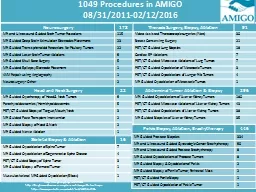
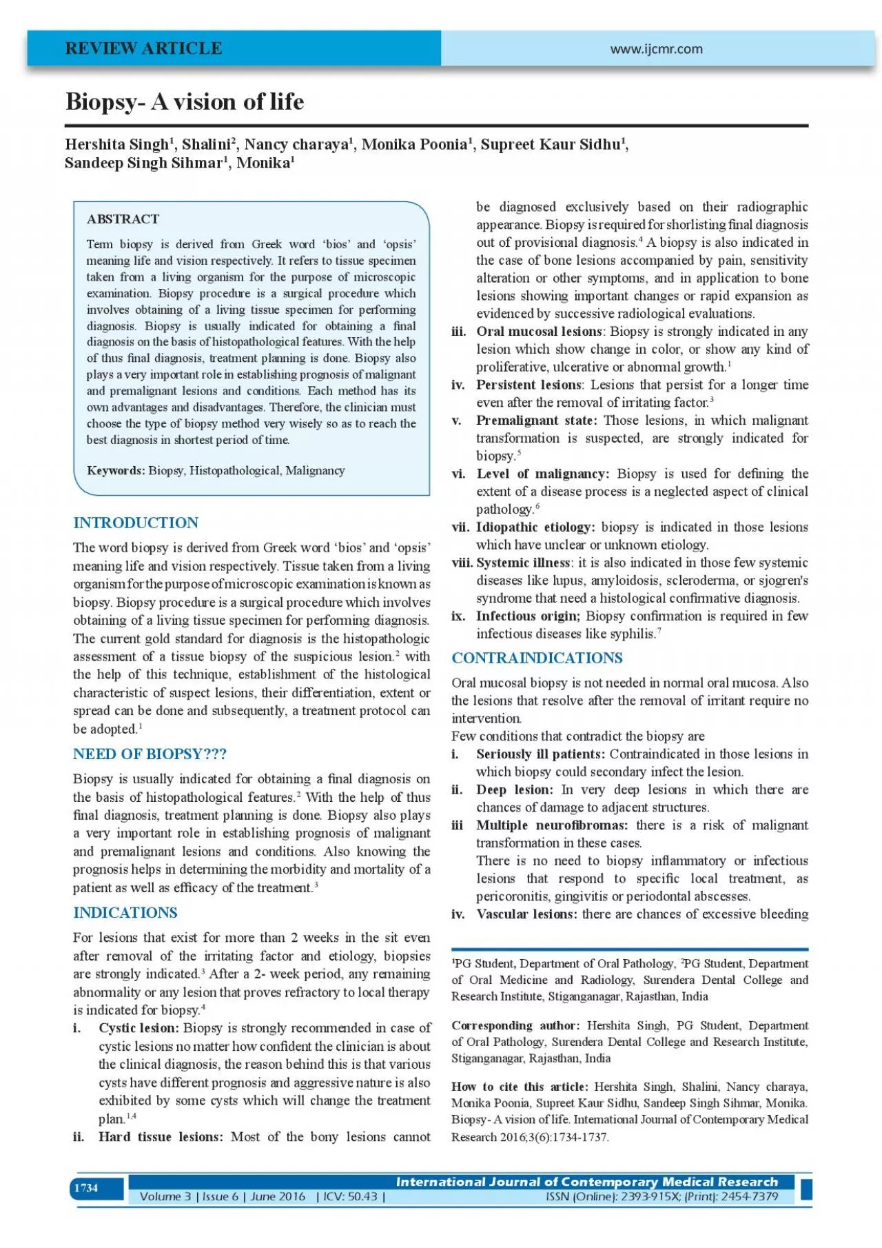
1734 Hershita Singh 1 Shalini 2 Nancy charaya 1 Monika Poonia 1 Supreet Kaur Sidhu 1 Sandeep Singh Sihmar 1 Monika 1 REVIEW ARTICLE Term biopsy is derived from Greek word 145bios146 ID: 937837

x00660069 biopsy oral lesions biopsy x00660069 lesions oral tissue diagnosis lesion specimen cases scalpel depending operative technique site advantages

Share:

Link:

Embed:

Download Presentation from below link

Download Pdf The PPT/PDF document "Biopsy A vision of life" is the property of its rightful owner. Permission is granted to download and print the materials on this web site for personal, non-commercial use only, and to display it on your personal computer provided you do not modify the materials and that you retain all copyright notices contained in the materials. By downloading content from our website, you accept the terms of this agreement.


Presentation Transcript

1734 Biopsy- A vision of life Hershita Singh 1 , Shalini 2 , Nancy charaya 1 , Monika Poonia 1 , Supreet Kaur Sidhu 1 , Sandeep Singh Sihmar 1 , Monika 1 REVIEW ARTICLE Term biopsy is derived from Greek word ‘bios’ and ‘opsis’ meaning life and vision respectively. It refers to tissue specimen taken from a living organism for the purpose of microscopic examination. Biopsy procedure is a surgical procedure which involves obtaining of a living tissue specimen for performing diagnosis. Biopsy is usually indicated for obtaining a �nal diagnosis on the basis of histopathological features. With the help of thus �nal diagnosis, treatment planning is done. Biopsy also INTRODUCTION The word biopsy is derived from Greek word ‘bios’ and ‘opsis’ meaning life and vision respectively. Tissue taken from a living organism for the purpose of microscopic examination is known as biopsy. Biopsy procedure is a surgical procedure which involves obtaining of a living tissue specimen for performing diagnosis. The current gold standard for diagnosis is the histopathologic assessment of a tissue biopsy of the suspicious lesion. 2 with the help of this technique, establishment of the histological characteristic of suspect lesions, their differentiation, extent or be adopted. 1 NEED OF BIOPSY??? Biopsy is usually indicated for obtaining a �nal diagnosis on the basis of histopathological features. 2 With the help of thus �nal diagnosis, treatment planning is done. Biopsy also plays a very important role in establishing prognosis of malignant and premalignant lesions and conditions. Also knowing the prognosis helps in determining the morbidity and mortality of a 3 INDICATIONS For lesions that exist for more than 2 weeks in the sit even after removal of the irritating factor and etiology, biopsies are strongly indicated. 3 abnormality or any lesion that proves refractory to local therapy is indicated for biopsy. 4 i.Cystic lesion: Biopsy is strongly recommended in case of cystic lesions no matter how con�dent the clinician is about the clinical diagnosis, the reason behind this is that various cysts have different prognosis and aggressive nature is also exhibited by some cysts which will change the treatment plan. ii.Hard tissue lesions: Most of the bony lesions cannot be diagnosed exclusively based on their radiographic appearance. Biopsy is required for shorlisting �nal diagnosis out of provisional diagnosis. 4 A biopsy is also indicated in alteration or other symptoms, and in application to bone lesions showing important changes or rapid expansion as evidenced by successive radiological evaluations. iii.Oral mucosal lesions : Biopsy is strongly indicated in any lesion which show change in color, or show any kind of 1 iv. : Lesions that persist for a longer time even after the removal of irritating factor. 3 v.Premalignant state: Those lesions, in which malignant transformation is suspected, are strongly indicated for biopsy. 5 Level of malignancy: extent of a disease process is a neglected aspect of clinical pathology. 6 vii.Idiopathic etiology: biopsy is indicated in those lesions which have unclear or unknown etiology. Systemic illness : it is also indicated in those few systemic diseases like lupus, amyloidosis, scleroderma, or sjogren's ix.Infectious origin; Biopsy con�rmation is required in few infectious diseases like syphilis. 7 CONTRAINDICATIONS Oral mucosal biopsy is not needed in normal oral mucosa. Also the lesions that resolve after the removal of irritant require no intervention. Few conditions that contradict the biopsy are Seriously ill patients: Contraindicated in those lesions in ii.Deep lesion: In very deep lesions in which there are iii Multipleneuro�bromas: there is a risk of malignant transformation in these cases. There is no need to biopsy in�ammatory or infectious lesions that respond to speci�c local treatment, as iv.Vascular lesions: there are chances of excessive bleeding 1 PG Student , Departmen

t of Oral Pathology, 2 PG Student, Department www.ijcmr.com International Journal of Contemporary Medical Research Volume 3 | Issue 6 | June 2016 | ICV: 50.43 |ISSN (Online): 2393-915X; (Print): 2454-7379 Research Institute, Stiganganagar, Rajasthan, India Corresponding author: Hershita Singh, PG Student, Department of Oral Pathology, Surendera Dental College and Research Institute, Stiganganagar, Rajasthan, India How to cite this article: Hershita Singh, Shalini, Nancy charaya, Monika Poonia, Supreet Kaur Sidhu, Sandeep Singh Sihmar, Monika. Biopsy- A vision of life. International Journal of Contemporary Medical 1735 in cases of vascular lesions. 7 The standard biopsy techniques may require modi�cation in some patient; including those with conditions that preclude the safe use of local anesthetic and those with severe bleeding diatheses or coagulopathies. 3 v.Esthetic reasons: Biopsy is contraindicated inn lesion in which biopsy can cause esthetic changes. 8 vi. Site Sites which are richly supplied by vasculature and in which there are chances of improper healing, biopsy should be done with great caution. Bisphosphonate therapy: Due to risk of development of osteoradionecrosis. 9 TYPES OF BIOPSY Biopsy reports, to a great extent are dependent upon the amount and the type of tissue specimen obtained from the lesion site. Depending upon location, depth and various other parameters, biopsy can by of various types. 4 Therefore; biopsy can be of following types as shown in Table-1 . INCISIONAL BIOPSY In order to make de�nitive diagnosis, removal of a representative sample of the lesion and normal adjacent tissue is done. If the lesion is extensive, different samples should be obtained, placing each of the in a separate and adequately identi�ed container. 1 Advantages 1.Only a small fragment of tissue is required. 11 Can be done in cases of suspected malignancy and premalignancy. In cases in which is dif�cult to excide the lesion due to large size. 1 It is also used in establishing the diagnosis systemic and autoimmune disease process If the lesion is ulcerated, the clinician should strive to include a portion of the adjacent intact epithelium in the specimen. Disadvantages Avoided in vascular cases as it may cause profuse bleeding. 9 EXCISIONAL BIOPSY Involves complete excision of the affected lesion for both the diagnostic and therapeutic purposes. This type of biopsy is mostly recommended in those cases in which the size of biopsy is small. 3 Advantages Complete removal of the lesion. 8 4 This is the ideal method of diagnosis of small melanomas 3 Disadvantages Dif�cult to perform in large lesions. Should be avoided in cases where a high grade malignancy is suspected. 9 SCALPEL BIOPSY Tissue sampling is most commonly done using a scalpel blade. Advantage In cases of oral mucosal lesion. Disadvantage Vague histopathological de�nition histological misinterpretation resulting in false negatives and false positive should be kept in mind while interpreting the results of scalpel biopsies. In case of extensive lesions, it should be avoided as it can lead to misdiagnosis. 11 Variants of scalpel Electro scalpel Laser scalpel PUNCH BIOPSY Punch biopsy is usually used as an alternative to incision biopsies for small lesion at an accessible site. The lateral tongue and buccal mucosa are appropriate sites for punch biopsy, as it must be feasible for device to approach the mucosal surface perpendicularly. 8 Advantages Rapid, simple, safe and inexpensive technique for obtaining a representative sample of most oral zones Good esthetic results due to better and fast wound healing. 1 The punch is able to obtain several samples at the same time, and at different points, and generates less patient anxiety than the conventional scalpel. 12 Depending on the character - istics of the target lesions Directsuper�cially, with easy access) in depth and is covered by normally appearing mucosa or Depending on the technique used Incisional Excisional Depending on the material employed A conventional scalpel

A punch Electro scalpels Co2 laser Depending on the processing of the sample Analyzed frozen Embedding in methacrylate Depending on the clinical timing Intra-operative Extra-operative Depending on the location of the target lesion The salivary gland Bone Lymph nodes Other head and neck tissues Depending on the purpose of the biopsy Diagnostic Experimental Depending on the time Pre-operative Intra-operative Post-operative Table-1: Types of biopsies 1736 produces fewer artifacts then the scalpel biopsy 2 Disadvantages In case of larger lesions, it should be avoided as intensely vascularized or innervated areas cannot be samples by this method. Not recommended in case of deep lesions and is limited to epithelial or super�cial mesenchymal target tissues. 1 Caution should be taken while biopsying areas which are near to normal anatomical structures. 8 B- FORCEP Bermejo developed this instrument for helping in measuring the depth of the samples to facilitate better sectioning. The forceps are equipped with two cusps- one with a window- to allow compression of the target tissue between them. The target zone is positioned exposed within the window, and compressive effect of the cusps allows us to work in an ischemic �eld within the window. Compression by the forceps causes the sectioned portion, freed from its peripheral connective tissue attachments, to propel from window. 13 FROZEN SECTIONS For rapid diagnosis during intra-operative period, the sampled material is processed without �xation, frozen with dry ice. 1 Frozen sections can be �xed, stained. and mounted for permanent reference. 6 A specimen processed in this manner is not satisfactory for detailed study of the cells, but it is valuable because it is quick and gives the surgeon immediate information regarding the malignancy of a piece of tissue. Advantages Differentiate between benign and malignant state and between type malignancies. Evaluate tissue margins for involvement by malignancy, e.g. basal cell carcinomas. Determine type of tissue, e.g. Differentiate lymphoid tissue from parathyroid gland. For generating reports during intra-operative sites. 14 Contraindications For extensive complex lesions. BRUSH BIOPSY It is a noninvasive method of evaluating oral mucosal lesions for cellular dysplasia and atypia. It is a three layer Trans-epithelial exfoliative cytology technique. 14 A brush biopsy was initially introduced for cervical smears in gynecological lesions and was later modi�ed for oral smears too. This technique demonstrated better cell spreading on the objective slides compared with smears obtained by using the conventional wooden spatula as well as an improvement in the cellular adequacy of the smears. Brush biopsy is strictly indicated for mass screening of suspected premalignancy and malignancy. 4 Advantages In contrast to exfoliative cytology, the brush biopsy collects cells from the full thickness of the oral epithelium. Non-invasive, chair side procedure, easy to perform and painless. Dysplasia can be ruled out Suspected cases of candidiasis can be rapidly con�rmed through oral cavity. 4 Disadvantages Cannot be used as a substitute for scalpel biopsy Signi�cant false �nding may be observed due to sampling error. 14 FINE NEEDLE BIOPSY Fine Needle biopsy (FNB) is a minimally invasive technique which is particularly suitable for those sensitive areas where an incisional biopsy is contraindicated or is not possible. Although it does not provide a de�nite type speci�c diagnosis, it is used in conjunction to the clinical and radiological �ndings to rapidly provide the best possible initial assessment on which management decisions can be based. Advantages Safe Inexpensive Accurate diagnosis Low risk of infections Disadvantages Possibility of false negative results Site precision is very important FNB should never be considered a replacement for or the cause of delay in open biopsy when it is indicated POINTS TO BE REMEMBERED ABOUT BIOPSY Site of application of Local Anesthesia (LA) s

olution: L.A should administered deeper in the tissue or area surrounding the biopsy site. Tissue artifacts may appear on microscopic examination if the L.A is given in the biopsy marked area. 4 Incision planning: All major vessels, nerves and other anatomical structures should be preserved while planning the incision. The incision should be of adequate depth to include the entire layer of epithelium and a signi�cant portion of the underlying connective tissue. 4 Surgical skill: The biopsy specimen should be handled with great care. A technique sensitive procedure will help attain a minimal artifact biopsy that in turn prevents dif�culty in diagnosing histopathologically. Intra operative artifacts may include pressing the sample with the tweezers, particularly if toothed, as may produce tissue tears and “pseudomicrocysts etc. Specimen transportation: While transporting the specimen to the histopathological laboratory, the specimen should be labeled properly with the patient's name, age, date of biopsy, and site of biopsy. The orientation of the specimen should be marked with the suture thread at different labels. the specimen should be delivered to pathologist immediately. Never put specimen on paper or in tubes with cotton plugs. cellulose �bers ruin microtome knives. 6 Fixative: The specimen should be transported in 10% 1737 formalin. 70% ethanol can also be used. Isopropyl or methyl alcohol, saline or distilled water should never be used as it may cause cellular deformation. 4 CONCLUSION Treatment of premalignant, malignant, benign or systemic is largely dependent upon the accurate diagnosis. And the fact of the matter is that the histopathological diagnosis remains the gold standard for the diagnosis. Several methods and types of biopsies have been tried over the past. Each method has its own advantages and disadvantages. Therefore, the clinician must choose the type of biopsy method very wisely so as to reach the best diagnosis in shortest period of time. REFERENCE 1. Mota-Ramirez A, Silvestre FJ, Simo JM. Oral biopsy in dental practice. Med Oral Patol Oral Cir Bucal. 2. Poh C.F, Ng Samson, Berean K.W., Williams P.M., Rosin M.P., Zhang L. Biopsy and histopathologic diagnosis of oral premalignant and malignant lesions. JCDA. 2008; 74:283-288. 3. Ephros H, James W.D. Oral Tissue Biopsy [Internet]. 2014 [update 2014 Aug 4; cited 2015 Apr 4]. Available from: 4. Rosebush M.S, Anderson K.M, Rawal S.Y, Mincer H.H, Rawal Y.B. The Oral Biopsy: Indications, Techniques and Special Considerations. Journal of the tennessee dental 5. Gandolfo S, Carbone M, Carrozzo M, Scamuzzi S. Biopsy technics in oral oncology: excisional or incisional biopsy? A critical review of the literature and the author personal 6. Moore E.G. The Importance of biopsy Procedure. J.A.M.A. 7. Oliver R.J, Sloan P., Pemberton M.N. Oral biopsies: methods andapplications. British dental Journal. 2007; 8. Avon L.S, Hagen B.E. Oral Soft Tissue Biopsy: An Overview. J Can Dent Assoc. 2012;78(c75). 9. Shklar G. The effect of manipulation and incision on experimental carcinoma of hamster buccal pouch. Cancer Mehrotra R, Gupta D. K., Exciting new advances in oral cancer diagnosis: avenues to early detection. 2011;28;3:33. 11. Kinsukawa J, Suefuji Y, Ryu F, Noguchi R, Iwamoto O, Kameyama T. Dissemination of cancer cells into circulation occurs by incisional biopsy of oral squamous 12. Eisen D. The oral mucosal punch biopsy. A report of 140 cases. Arch Dermatol. 1992;128:815-7. 13. Bermejo FA, Lopez-J P. Instrument for biopsy of oral lesions: an improved chalazion forceps. Dermatol Surg. 14. Hall D.L. Oral Brush Biopsy Technique Instruction outcome for senior Dental Students. J dent Educ. 2006;4; Source of Support: Interest: Singh, et al.Biopsy International Journal of Contemporary Medical Research ISSN (Online): 2393-915X; (Print): 2454-7379 | ICV: 50.43 |Volume 3 | Issue 6 | June 2016 Singh, et al.Biopsy International Journal of Contemporary Medical Research Volume 3 | Issue 6 | June 2016 | ICV: 50.43 |ISSN (Online): 2393-915X; (Print): 2454-737