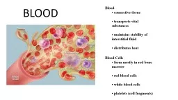
transports maintains stability of distributes Blood Cells form mostly in blood cells blood cells ID: 912854
Download Presentation The PPT/PDF document "BLOOD Blood __________________" is the property of its rightful owner. Permission is granted to download and print the materials on this web site for personal, non-commercial use only, and to display it on your personal computer provided you do not modify the materials and that you retain all copyright notices contained in the materials. By downloading content from our website, you accept the terms of this agreement.
Slide1
BLOOD
Blood
__________________ transports ________ maintains stability of _________________ distributes __________
Blood Cells
form mostly in
_________
______blood
cells
_______blood
cells
_________(
cell fragments)
Slide2Blood
_________
___________RBC(__________)WBC(_________)Platelets(__________)_____
Contains:
_______________
_____________
_____________________
______________
_________
______________
_______________
___________________________
__________________
Immunity: B-cells, T-cells
_________________
_______________
Slide3Slide4Slide5Slide6Erythrocyte: 400x
Neutrophil: 400x
Basophil: 400xEosinophil: 400xLymphocyte: 400xMonocyte: 400x
Slide7Slide8Name
Size
Diff. %Granular (Y or N)Event of IncreaseBLOOD CELL REVIEW
Slide9Slide10Slide11BLOOD
Blood
connective tissue transports vital substances maintains stability of interstitial fluid distributes heat
Blood Cells
form mostly in red bone marrow
red blood cells
white blood cells
platelets (cell fragments)
Slide12Blood
Cellular
Non-CellularRBC(erythrocytes)WBC(Leukocytes)Platelets(thrombocytes)PlasmaNeutrophils
Basophils
Monocytes
Lymphocytes
Eosinophils
Granulocytes
Agranulocytes
Contains
:
Electrolytes
Gases
Water
Proteins
Nutrients
Waste
Phagocytosis
Bacterial infection
Worm infestations (parasites)
Allergic reactions
Injury
Release Heparin
(blood thinner)
Release Histamine
(inflammation)
Large phagocytic cell – mature into “Macrophages”
Infections from large bacteria and fungus,
dead cells, debris
Immunity: B-cells, T-cells
Viral infections
Transport O
2
and CO
2
Blood clotting
Slide13Slide14Slide1560%
2
%1%30%7%
Slide1660%
2%
<1%30%7%
Slide1717
Red Blood Cell Counts
number of RBCs in a cubic millimeter of blood
4,600,000 – 6,200,000 in males
4,200,000 – 5,400,000 in adult females
4,500,000 – 5,100,000 in children
reflects blood’s oxygen carrying capacity
Question: How can changes in these affect the health of the individual ranges?
Answ
: Increase O2 carrying capacity
!
Slide1818
White Blood Cell Counts
procedure used to count number of WBCs per cubic millimeter of blood 5,000 – 10,000 per cubic millimeter of blood
leukopenia
low WBC count (below 5,000)
typhoid fever, flu, measles, mumps, chicken pox, AIDS
leukocytosis
high WBC count (above 10,000)
acute infections, vigorous exercise, great loss of body fluids
differential WBC count
lists percentages of types of leukocytes
may change in particular diseases
Slide1919
Blood Platelets
thrombocytes cell fragments of megakaryocytes 130,000 – 360,000 per cubic millimeter of blood
helps control blood loss from broken vessels
Slide20Slide2121
straw colored
liquid portion of blood
55% of blood
92% water
Blood Plasma
Slide22The neutrophil are the more common leukocytes. They have a diameter of 12-15 µm. You can recognize them as their nucleus is divided into 2 - 5 lobes connected by a fine nuclear strand or filament (fig. 8). The cytoplasm is transparent because its granules are small and faintly pink colored. Immature neutrophils have a band-shaped or horseshoe-shaped nucleus and are known as band cells. In the nucleus of the neutrophil of cells from females, you may see an appendage like a little drumstick (Barr body). It is the second X chromosome, inactivated.
Slide23The eosinophils are quite rare in the blood. They have the same size as the neutrophils. Generally their nucleus is bi-lobed. But even nuclei with three or four lobes have been observed. The cytoplasm is full of granules which assume a characteristic pink-orange color (fig. 9). As for the neutrophil, the nucleus is still easily visible
Slide24Basophils are the rarest leukocytes: less than 1 %. They are quite small: 9-10 µm in diameter. Cytoplasm is very rich in granules which take a dark purple color. The nucleus is bi- or tri-lobed, but it is hard to see because of the number of granules which hide it (fig. 10).
Slide25Lymphocytes are quite common in the blood: 20-40%, 8-10 µm in diameter and generally they are smaller than the other leukocytes but they are still a few larger than red cells (fig. 11). The cytoplasm is transparent. The nucleus is round and large in comparison to the cell and it occupies most of it. In any case, some of the cytoplasm remains visible, generally in a lateral position. According to the quantity of cytoplasm, lymphocytes are divided into small, medium and large. With Giemsa stain, we cannot distinguish the different types of lymphocyte (B, T, NK), either in the blood because they are not activated, or because it would be necessary to perform special immunochemical staining
Slide26Monocytes are the biggest leukocytes: 16-20 µm. They have a great
reniform (kidney-shaped) or horseshoe-shaped nucleus, in some cases even bi-lobed. The cytoplasm is transparent, but with an appearance of "ground glass" (fig. 12).
Slide27Slide28Slide29Slide30Blood Types
Slide31Snake Venom