/
Con30ict of interests NoneSechi G Serra A Wernicke146s enceph Con30ict of interests NoneSechi G Serra A Wernicke146s enceph

Con30ict of interests NoneSechi G Serra A Wernicke146s enceph - PDF document

holly
holly . @holly
Follow
343 views
Uploaded On 2022-10-28

Con30ict of interests NoneSechi G Serra A Wernicke146s enceph - PPT Presentation

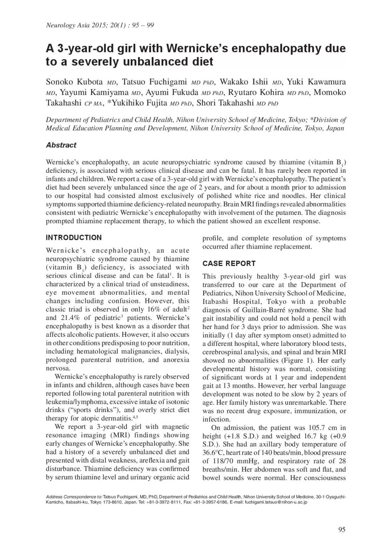
99 Neurology AsiaMarch 2015 myelin Coexistence of polyneuropathy may cause some patients with Wernicke146s encephalopathy to experience limb ataxia and dysarthria It is estimated that 19 of pati ID: 961574

wernicke 146 thiamine encephalopathy 146 wernicke encephalopathy thiamine patient diet clinical brain showed mri ciency autism admission putamen pediatric

Share:

Link:

Embed:

Download Presentation from below link

Download Pdf The PPT/PDF document "Con30ict of interests NoneSechi G Serra ..." is the property of its rightful owner. Permission is granted to download and print the materials on this web site for personal, non-commercial use only, and to display it on your personal computer provided you do not modify the materials and that you retain all copyright notices contained in the materials. By downloading content from our website, you accept the terms of this agreement.


Presentation Transcript

99 Conict of interests: NoneSechi G, Serra A. Wernicke’s encephalopathy: new clinical settings and recent advances in diagnosis and management. Lancet Neurol 2007; 6:442-55Harper CG, Giles M, Finlay-Jones R. Clinical signs in the Wernicke-Korsakoff complex: a retrospective analysis of 131 cases diagnosed at necropsy. J Neurol Neurosurg Psychiatry 1986; 49:341-53.Vasconcelos MM, Silva KP, Vidal G, Silva AF, Domingues RC, Berditchevsky CR. Early diagnosis of pediatric Wernicke’s encephalopathy. Pediatr Neurol 1999; 20:289-944.Svahn J, Schiafno MC, Caruso U, Calvillo M, Minniti G, Dufour C. Severe lactic acidosis due to thiamine deciency in a patient with B-cell leukemia/lymphoma on total parenteral nutrition during high-dose methotrexate therapy. J Pediatr Hematol Oncol2003; 25:965-8.5.Saeki K, Saito Y, Komaki H, et al. Thiamine-decient encephalopathy due to excessive intake of isotonic drink or overstrict diet therapy in Japanese children. Brain Dev 2010; 32:556-63.6.Torvik A, Lindboe CF, Rogde S. Brain lesions in alcoholics: a neuropathological study with clinical J Neurol Sci 1982; 56:233-487.Sechi GP, Bosincu L, Cossu Rocca P, Deiana GA, Correddu P, Murrighile RM. Hyperthermia, choreic dyskinesias and increased motor tone in Wernicke’s encephalopathy. Eur J Neurol 1996; 3(Suppl Victor M, Adams RD, Collins GH. The Wernicke-Korsakoff syndrome: a clinical and pathological study of 245 patients, 82 with post-mortem examinations. Contemp Neurol Ser 1971; 7: 1-206.9.Tanphaichitr V. Thiamine. In: Shils ME, Olson JA, Shike M, Ross AC, eds: Modern nutrition in health and disease, 9th ed. Baltimore: Williams and Wilkins, 10.Koguchi K, Nakatsuji Y, Abe K, Sakoda S. Wernicke’s encephalopathy after glucose infusion. Neurology2004; 62:512.11.Antunez E, Estruch R, Cardenal C, Nicolas JM, Fernandez-Sola J, Urbano-Marquez A. Usefulness of CT and MR imaging in the diagnosis of acute Wernicke’s encephalopathy. AJR Am J Roentgenol1998; 171:1131-7.12.Chung SP, Kim SW, Yoo IS, Lim YS, Lee G. Magnetic resonance imaging as a diagnostic adjunct to Wernicke’s encephalopathy in the ED. Am J Emerg 2003; 21:497-502.Mascalchi M, Simonelli P, Tessa C, et al. Do acute lesions of Wernicke’s encephalopathy show contrast enhancement? Report of three cases and review of Neuroradiology 1999; 41:249-54.Zuccoli G, Siddiqui N, Bailey A, Bartoletti SC. Neuroimaging findings in pediatric Wernicke encephalopathy: a review. Neuroradiology 2010; 15.Field D, Garland M, Williams K. Correlates of specic childhood feeding problems. J Paediatr Child Health2003; 39:299-304.16.Schreck KM, Williams K, Smith AF. A comparison of eating behaviors betweenchildren with and without J Autism Devel Disord 2004; 34: 433-8.17.Gillberg C, Billstedt E. Autism and Asperger syndrome: coexistence with other other clinical Acta Psychiatr Scand 2000; 102: 321-30.18.Watanabe S, Yamakura S, Hirano K, Okumura Y, Aiba H. A case of infantile autism with pediatric Wernicke’s encephalopathy due to severe eating disorder (in Japanese, abstract English). No to Hattatsu 2009; Neurology AsiaMarch 2015 myelin. Coexistence of polyneuropathy may cause some patients with Wernicke’s encephalopathy to experience limb ataxia and dysarthria. It is estimated that 19% of patients with Wernicke’s encephalopathy present with none of the classic , although at least one symptom will usually appear during the course of the Early diagnosis of Wernicke’s encephalopathy and beriberi can result in the successful reversal

of many of the neurological abnormalities with simple thiamine replacement therapy. However, once Korsakoff’s dementia develops in the later stages, the recovery rate is much lower, and the estimated mortality is 17%. Fortunately, our patient appears to have recovered fully. We speculated that our patient had early and mild Wernicke’s encephalopathy.Because the body’s reserves of thiamine are sufcient for up to 18 days, in a healthy individual, any condition of unbalanced nutrition that lasts 2–3 weeks may lead to Wernicke’s encephalopathy. In individuals with marginal stores of thiamine, the disorder may occur earlier, particularly if the diet has been very rich in carbohydrates. About two-thirds of the world’s population has rice as the main staple of their diet. Polished white rice is highly decient in thiamine because milling removes the husk, which contains most of the thiamine. Our patient had eaten a severely unbalanced diet from the age of 2 years. Following the birth of her sister four months prior to admission to our hospital, her diet consisted almost exclusively of polished white rice and noodles because her mother no longer had time to cook for her. The daily thiamine requirement is related to energy need and increases with carbohydrate intake, infection, and physical activity. In this patient, increased carbohydrate intake resulted in relative thiamine deciency.MRI is currently the best method for conrming a clinical suspicion of Wernicke’s encephalopathy, owing to its high specicity of 93%. MRI studies typically show a bilaterally symmetrical increase in T2 signal in the paraventricular regions of the thalamus, hypothalamus, mammillary bodies, periaqueductal region, oor of the fourth ventricle, and midline cerebellum. The patient’s brain MRI displayed abnormalities consisting of increased signal T2-weighted and FLAIR images of bilateral lesions in the putamen and pons, and a lesion in the cerebral aqueduct. In a review of 23 cases with pediatric Wernicke’s encephalopathy, 16 cases showed bilateral alterations in the thalamus (70%), 13 in the periaqueductal region (57%), 11 in the mammillary bodies (47%), and 9 in putamen (39%). Symmetric basal ganglia alterations with involvement of the putamen have only been observed in children. Basal ganglia alterations with involvement of the putamen appear to differentiate the pediatric form of Wernicke’s encephalopathy from the adult cases. To explain the selective involvement of the basal ganglia (specically, alterations in the putamen) in pediatric patients with Wernicke’s encephalopathy, et al. speculated that thiamine-dependent metabolism is increased during development.In the case reported here, clinical features such as claw hand and foot drop supported thiamine deciency-related neuropathy. However, brain MRI ndings revealed abnormalities consistent with Wernicke’s encephalopathy. We therefore believe that this case may represent very early stage pediatric Wernicke’s encephalopathy without the appearance of the classic triad. The MRI was useful for this early diagnosis.Findings from developmental psychological studies of this patient (Tanaka-Binet Intelligence Quotient test, Goodenough-Harris Draw-a-Person intelligence test, PARS score, and Theory of Mind Development Screening Test), indicated autism spectrum disorder as a possible underlying disorder. Sixty-two percent of children with autism present with food selectivity by type.Childre

n with autism have signicantly more feeding problems and eat a narrower range of foods than children without autism. Pica, overeating, anorectic behavior, complete food refusal, as well as unusual food restriction and preferences are recognized in autism. Watanabe et al. reported a case of a 3-year-old boy with infantile autism and a severe eating disorder who developed Wernicke’s encephalopathy.The ndings in our patient demonstrated that Wernicke’s encephalopathy should be considered when patients with severe eating disorders present with neurological disturbance.In conclusion, we report a 3-year-old girl in whom brain MRI showed very early changes of Wernicke’s encephalopathy, and biochemical testing conrmed thiamine deciency. The patient had eaten a severely unbalanced diet from the age of 2 years, consisting almost entirely of polished white rice and noodles from about a month prior to admission to our hospital. This prompted initiation of thiamine replacement therapy, to which the patient showed an excellent response. Her severely unbalanced diet was suspected to be a result of a feeding abnormality due to autistic spectrum disorder. 97 and brainstem auditory evoked potentials showed no abnormal ndings. Motor nerve conduction velocity (NCV) was performed on the 3rd day after admission. No M or F wave responses were obtained at the right median or right tibial nerves. Sensory NCV showed no response at the right sural nerve. Needle electromyography was not performed because the patient was very young. There were no abnormal ndings on her electrocardiography or echocardiography.The patient had consumed a severely unbalanced diet from the age of 2 years, consisting mainly of polished white rice and noodles. Serum thiamine levels were low (24 ng/mL; normal, 24–66 ng/mL). Wernicke’s encephalopathy was diagnosed, and the patient was given 100 mg/day of intravenous thiamine for 7 days, from the 4th day after admission. This was followed by oral thiamine (100 mg/day). Her mother was instructed on how to implement a balanced diet. On the 17th day after admission, she was discharged from the hospital without any neurological abnormality. Upon discharge, her urinary organic acid prole was normal. Her motor NCV at the left and right tibial nerves had improved but was still delayed (left: 30.6 m/s, right: 33.8 m/s).During her hospitalization, the patient often had difculty understanding verbal instructions. It was also noted that she easily became frustrated with her mother. Her intelligence score, using the Tanaka-Binet Intelligence Quotient, was normal at 115 (chronological age: 3 years and 11 months, mental age: 4 years and 6 months). However, in the Goodenough-Harris Draw-a-Person intelligence test, her score was 85.The Pervasive Developmental Disorders-Autism Society Japan Rating Scale (PARS) is Figure 3.Brain magnetic resonance imaging. T2-weighted uid-attenuated inversion recovery image one month after discharge showing complete resolution of the lesions. a screening test for behavioral problems and pervasive developmental disorders (PDD). The scale comprises 57 questions, assessing eight different characteristic domains of children with PDD. The patient’s PARS score was 11 points (PDD is strongly suggested at 9 or more points). In the Theory of Mind Development Screening Test, she did not pass the standard false-belief task, often called the “Sally-Anne test” (usually passed at aged 4 or 5 years). She also failed the facial

expression recognition task (usually passed at 3 years). These ndings suggested autistic spectrum disorder. A follow-up brain MRI 1 month after discharge revealed complete resolution of lesions in the putamen, pons, and cerebral aqueduct (Figure 3). Motor NCV at the right tibial nerve had improved further but was still slightly delayed (36.8 m/s). Oral thiamine therapy was discontinued approximately 2 months after discharge. When the patient was seen a month later, she was eating a balanced diet and had no abnormal clinical symptoms. Her serum thiamine level was normal at 57 ng/mL.Thiamine (vitamin B) deciency results in a spectrum of complications including peripheral neuropathy (dry beriberi), cardiomyopathy (wet beriberi), and Wernicke’s encephalopathy. Several factors, such as genetic susceptibility and age, can inuence the symptoms of thiamine deciency. While acute severe deciency of thiamine usually causes Wernicke’s encephalopathy, a chronic deciency may result in peripheral neuropathy, which is often distal and preferentially affects Neurology AsiaMarch 2015 was normal. She could raise her arms but had a clawed hand posture and could not hold objects. She could maintain a sitting, but not standing, position. She had bilateral foot drop and gait was difcult. Her muscle power was grade 3–4 in her upper and lower extremities. Deep tendon reexes were impaired in her arms and legs, but she had no loss of sensation to pain or temperature. She did not have nystagmus, hand tremors, or obvious truncal ataxia.Laboratory blood tests were normal for full blood count, serum electrolytes, total protein, albumin, blood urea nitrogen, creatinine, aspartate aminotransferase, alanine aminotransferase, lactate dehydrogenase, alkaline phosphatase, creatine kinase, glucose, and C-reactive protein. Figure 1.Brain magnetic resonance imaging. T2 weighted uid-attenuated inversion recovery image taken at a different hospital 2 days before admission to our hospital with no abnormal ndings. Figure 2.Brain magnetic resonance imaging. T2-weighted uid-attenuated inversion recovery image on the 2nd day of admission showed bilateral lesions in the putamen and pons, and a lesion in the cerebral Cerebrospinal uid (CSF) showed no pleocytosis, and normal protein and sugar. Myelin basic protein and oligoclonal immunoglobulin G in CSF were negative. Lactate/pyruvate levels were 19.1/1.18 mg/dL in the blood and 12.8/0.79 mg/dL in the CSF.The urinary organic acid prole showed massive excretion of lactate with high levels of pyruvate, increased excretion of 3-OH-butyrate and acetoacetate, mild excretion of alpha-ketoglutarate and oxaloacetate, and massive excretion of palmitate. A brain MRI on T2-weighted uid-attenuated inversion recovery (FLAIR) sequences showed hyperintensity lesions bilaterally in the putamen and pons, and a lesion in the cerebral aqueduct (Figure 2). Her electroencephalography A 3-year-old girl with Wernicke’s encephalopathy due to a severely unbalanced dietSonoko Kubota , Tatsuo Fuchigami MD PhD, Wakako Ishii , Yuki Kawamura , Yayumi Kamiyama , Ayumi Fukuda MD PhD, Ryutaro Kohira MD PhD, Momoko Takahashi CP MA, *Yukihiko Fujita MD PhD, Shori Takahashi MD PhDDepartment of Pediatrics and Child Health, Nihon University School of Medicine, Tokyo; *Division of Medical Education Planning and Development, Nihon University School of Medicine, Tokyo, JapanAbstract Wernicke’s encephalopathy, an acute neuropsychiatric syndrome caus

ed by thiamine (vitamin Bdeciency, is associated with serious clinical disease and can be fatal. It has rarely been reported in infants and children. We report a case of a 3-year-old girl with Wernicke’s encephalopathy. The patient’s diet had been severely unbalanced since the age of 2 years, and for about a month prior to admission to our hospital had consisted almost exclusively of polished white rice and noodles. Her clinical symptoms supported thiamine deciency-related neuropathy. Brain MRI ndings revealed abnormalities consistent with pediatric Wernicke’s encephalopathy with involvement of the putamen. The diagnosis prompted thiamine replacement therapy, to which the patient showed an excellent response.Neurology Asia 2015; 20(1) : 95 – 99Address Correspondence to: Tatsuo Fuchigami, MD, PhD, Department of Pediatrics and Child Health, Nihon University School of Medicine, 30-1 Oyaguchi-Kamicho, Itabashi-ku, Tokyo 173-8610, Japan. Tel: +81-3-3972-8111, Fax: +81-3-3957-6186, E-mail: fuchigami.tatsuo@nihon-u.ac.jWernicke’s encephalopathy, an acute neuropsychiatric syndrome caused by thiamine (vitamin B) deficiency, is associated with serious clinical disease and can be fatal. It is characterized by a clinical triad of unsteadiness, eye movement abnormalities, and mental changes including confusion. However, this classic triad is observed in only 16% of adultand 21.4% of pediatric patients. Wernicke’s encephalopathy is best known as a disorder that affects alcoholic patients. However, it also occurs in other conditions predisposing to poor nutrition, including hematological malignancies, dialysis, prolonged parenteral nutrition, and anorexia nervosa.Wernicke’s encephalopathy is rarely observed in infants and children, although cases have been reported following total parenteral nutrition with leukemia/lymphoma, excessive intake of isotonic drinks (“sports drinks”), and overly strict diet therapy for atopic dermatitis.We report a 3-year-old girl with magnetic resonance imaging (MRI) findings showing early changes of Wernicke’s encephalopathy. She had a history of a severely unbalanced diet and presented with distal weakness, areexia and gait disturbance. Thiamine deciency was conrmed by serum thiamine level and urinary organic acid prole, and complete resolution of symptoms occurred after thiamine replacement. CASE REPORTThis previously healthy 3-year-old girl was transferred to our care at the Department of Pediatrics, Nihon University School of Medicine, Itabashi Hospital, Tokyo with a probable diagnosis of Guillain-Barré syndrome. She had gait instability and could not hold a pencil with her hand for 3 days prior to admission. She was initially (1 day after symptom onset) admitted to a different hospital, where laboratory blood tests, cerebrospinal analysis, and spinal and brain MRI showed no abnormalities (Figure 1). Her early developmental history was normal, consisting of signicant words at 1 year and independent gait at 13 months. However, her verbal language development was noted to be slow by 2 years of age. Her family history was unremarkable. There was no recent drug exposure, immunization, or On admission, the patient was 105.7 cm in height (+1.8 S.D.) and weighed 16.7 kg (+0.9 S.D.). She had an axillary body temperature of 36.6°C, heart rate of 140 beats/min, blood pressure of 118/70 mmHg, and respiratory rate of 28 breaths/min. Her abdomen was soft and at, and bowel sounds were normal. Her conscious