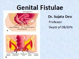
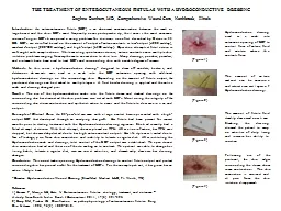
Mr Darren TONKIN MBBS FRACS The Queen Elizabeth Hospital Adelaide SA Background Enterocutaneous fistulae abnormal connection between GI tract and skin Majority gt75 develop postoperatively ID: 786681
Download The PPT/PDF document "The Management of Enterocutaneous Fistul..." is the property of its rightful owner. Permission is granted to download and print the materials on this web site for personal, non-commercial use only, and to display it on your personal computer provided you do not modify the materials and that you retain all copyright notices contained in the materials. By downloading content from our website, you accept the terms of this agreement.
Slide1
The Management of Enterocutaneous Fistulae
Mr Darren TONKIN (MBBS, FRACS)The Queen Elizabeth HospitalAdelaide, SA
Slide2Slide3Slide4Background
Enterocutaneous fistulae = abnormal connection between GI tract and skinMajority (>75%) develop postoperatively
1
Malignancy
IBD
Intra-abdominal sepsis
Dense adhesions
Open abdomen
Remainder spontaneous2IBD (esp Crohn’s)Radiation enteritisDiverticular diseaseMalignancyTraumaIntra-abdominal sepsis
Berry SM, Fischer JE. Classification and pathophysiology of enterocutaneous fistulas. Surg Clin North Am. 1996;76:1009-1018.
Hollington P, Mawdsley J, Lim W, Gabe SM, Forbes A, Windsor AJ. An 11-year experience of enterocutaneous fistula. Br J Surg. 2004;91:1646-1651.
Slide5Background
Complex patients with considerable morbidity and mortalityMortality rates dropped from ~60% in ‘60’s1
to less than 10%
2,3
Operative mortality <5%
3
Multidisciplinary approach
Spontaneous closure 7-70% reported
2,3 depends on aetiology & referral patternPATIENCE!Edmunds LH Jr, Williams GM, Welch CE. External fistulas arising from the gastro-intestinal tract. Ann Surg 1960;152:445-471.
Hollington P, Mawdsley J, Lim W, Gabe SM, Forbes A, Windsor AJ. An 11-year experience of enterocutaneous fistula. Br J Surg. 2004;91:1646-1651.
Datta V, Engledow A, Chan S, Forbes A, Cohen CR, Windsor A. The management of enterocutaneous fistula in a regional unit in the United Kingdom: A prospective study. Dis Colon Rectum 2010;53:192-199.
Slide6Classification
SiteSmall bowel (65%)Colon (30%)Stomach/oesophagus (rare)
Output
Low (<200 mL/24 hr)
Moderate (200 – 500 mL/24 hr)
High (>500 mL/24 hr)
Complexity
Simple
Complex – long, multiple, associated abscess, other organ involvement (e.g. bladder, vagina)
Slide7Management
Multidisciplinary approachSurgeonPhysicianDietician
Pharmacist
Stomal therapist
Radiologist
Social worker
Slide8Steps in Management
ResuscitationElimination of sepsisWound managementOptimisation of nutrition
Assessment of anatomy
Definitive surgery
Slide9Resuscitation
Correction of fluid and electrolyte imbalancesOpen abdomen is equivalent to large full thickness burn in terms of fluid lossesElectrolyte replacement (esp. Na+
, K
+
, Mg
2+
)
Close monitoring of input and output
IDC, measure stoma losses, CVP measurement
Slide10Control Sepsis
Sepsis is most common cause of mortality in ECF patients (approx 2/3)1CT scan
percutaneous drainage of intra-abdominal abscesses
open drainage if superficial
Rarely laparotomy if peritonitis present
exteriorise fistula or proximal diversion
Line associated sepsis not be overlooked (more common with central line vs. PICC)
2
Hollington P, Mawdsley J, Lim W, Gabe SM, Forbes A, Windsor AJ. An 11-year experience of enterocutaneous fistula. Br J Surg. 2004;91:1646-1651.
Collignon, PJ. Intravascular catheter associated sepsis: a common problem. The Australian Study on Intravascular Catheter Associated Sepsis. Med J Aust
.
1994 Sep 19;161(6):374-8.
Slide11Wound Management
Protect skin from corrosive fistula effluentCareful measurement of fistula outputExperienced stomal therapist essential“creative bagging”
Open abdomen
vacuum dressings with isolation of fistula and protection of other bowel segments
don’t apply sponge directly to bowel or anastomosis
Slide12Open Abdomen
Bogota bag
Vacuum dressing
Slide13Slide14Nutrition
Aim to return malnourished patient to health, allowing spontaneous closure or optimisation for future surgeryEnteral vs. TPN debate (no L1 evidence)
Enteral preferred
Avoids line-related complications (sepsis, thrombosis, pneumothorax)
Trophic effect on bowel mucosa
1
Supports immunological, barrier and hormonal functions of gut
2High calorie, protein supplements, electrolyte mix, minimise hypotonic fluids and drinking with mealsFistuloclysis may be used to avoid TPN3
Datta V, Engledow A, Chan S, Forbes A, Cohen CR, Windsor A. The management of enterocutaneous fistula in a regional unit in the United Kingdom: A prospective study. Dis Colon Rectum 2010;53:192-199.
Schecter, WP, Hirshberg A, Chang DS, Harris HW, Napolitano LM, Wexner SD, Dudrick SJ. Enteric fistulas: Principles of management. J Am Coll Surg 2009;209: 484-491.
Tuebner A, Morrison K, Ravishankar HR, Anderson ID, Scott NA, Carlson GL. Fistuloclysis can successfully replace parenteral feeding in the nutritional support of patients with enterocutaneous fistula. Br J Surg 2004;91:625-631.
Slide15Nutrition
TPN may be required if high output, distal obstruction, ongoing sepsisHigh dose anti-diarrhoeals (loperamide, codeine), proton pump inhibitor
1,2
Octreotide & somatostatin
can reduce fistula output and time to spontaneous closure
3
no evidence for improved closure rate
4
expensivenot routinely usedDatta V, Engledow A, Chan S, Forbes A, Cohen CR, Windsor A. The management of enterocutaneous fistula in a regional unit in the United Kingdom: A prospective study. Dis Colon Rectum 2010;53:192-199.
Hollington P, Mawdsley J, Lim W, Gabe SM, Forbes A, Windsor AJ. An 11-year experience of enterocutaneous fistula. Br J Surg. 2004;91:1646-1651.
Dorta G. Role of octreotide and somatostatin in the treatment of intestinal fistulae. Digestion 1999;60:53-56.
Alivizatos V, Felekis D, Zorbalas A. Evaluation of the effectiveness of octreotide in the conservative management of postoperative enterocutaneous fistulas. Hepatogastroenterology 2002;49:1010-1012.
Slide16Spontaneous Closure
May occur during the “waiting period”7-70% spontaneous closure reported1,2
, varies with referral patterns and underlying cause
90% within 1
st
month, none after 3 months
Octreotide may speed closure
3
, no improvement in closure rate4Hollington P, Mawdsley J, Lim W, Gabe SM, Forbes A, Windsor AJ. An 11-year experience of enterocutaneous fistula. Br J Surg. 2004;91:1646-1651.
Datta V, Engledow A, Chan S, Forbes A, Cohen CR, Windsor A. The management of enterocutaneous fistula in a regional unit in the United Kingdom: A prospective study. Dis Colon Rectum 2010;53:192-199.
Dorta G. Role of octreotide and somatostatin in the treatment of intestinal fistulae. Digestion 1999;60:53-56.
Alivizatos V, Felekis D, Zorbalas A. Evaluation of the effectiveness of octreotide in the conservative management of postoperative enterocutaneous fistulas. Hepatogastroenterology 2002;49:1010-1012.
Slide17Spontaneous Closure
Favourable
Unfavourable
Long tract
Short, wide
tract, e
version of mucosa
Intestinal continuity
Disruption
of GIT
No distal obstruction
Distal obstruction
No sepsis
Sepsis
Low
output
High output
Good nutrition
Malnutrition
No
underlying bowel disease
Disease
d bowel (egg Crohn’s, malignancy, radiation enteritis)
Slide18Assess Anatomy
CT – fistula + abdominal wallContrast studies - roadmap
Slide19Definitive Surgery
Wait at least 3 months for resolution of obliterative peritonitis.Most centres recommend >6 month wait from last laparotomy
1
Signs that adhesions have “matured” (neoperitoneum formed)
1
Fistulae prolapse
Skin/skin graft loose over bowel
Ensure whole day list available, ICU available postop
Hollington P, Mawdsley J, Lim W, Gabe SM, Forbes A, Windsor AJ. An 11-year experience of enterocutaneous fistula. Br J Surg. 2004;91:1646-1651.
Slide20Technique
Full adhesiolysis, including laterallyAvoid enterotomies, repair immediately if occurResection of fistulating segment with preservation of as much enteric length as possible
Measure residual length carefully (ruler and tape)
Resection and anastomosis preferred over fistula closure (>35% recurrence with simple closure
1
)
Defunctioning stoma’s as needed
Abdominal wall reconstruction a major challenge
Success rateApprox 60% with simple fistula closure1>80% with formal resection1,2More than one procedure may be required2
Higher recurrence with Crohn’s, irradiation etc
1
Lynch AC, Delaney CP, Senagore AJ, Connor JT, Remzi FH, Fazio VW. Clinical outcome and factors predictive of recurrence after enterocutaneous fistula surgery. Ann Surg 2004;240:825-31
Hollington P, Mawdsley J, Lim W, Gabe SM, Forbes A, Windsor AJ. An 11-year experience of enterocutaneous fistula. Br J Surg. 2004;91:1646-1651.
Slide21Abdominal Wall Reconstruction
Fascial edges often retracted widely (open abdomen)Avoid synthetic meshDissolvable meshBiologic mesh
Component separation technique
Avoid open abdomen (increased risk of re-fistulation)
Slide22Abdominal Wall
Loss of domain
Enterocutaneous fistula
Slide23Components Separation
Ramirez 1990 – cadaveric and 11 patients1Incise external oblique aponeurosis and mobilize in plane deep to EO, incise rectus sheath and separate rectus from posterior sheath
Allows medialisation of rectus, to obtain midline fascial closure
8+2cm advancement at umbilicus (each side)
Ramirez OM, Ruas E, Dellon AL. “Components Separation” method for closure of abdominal-wall defects: an anatomic and clinical study. Plastic and Reconstructive Surgery. 1990;86:519-526.
Slide24Technique
Slide25Technique
Slide26Results
Up to 30% recurrence (no mesh)Approx 30 to 40% wound complicationsInfectionSeroma
Skin flap necrosis
Mesh reduces recurrence to 5 to 10%
Binder reduces seroma, but not recurrence (concern re flap ischaemia)
Slide27Mesh??
Mesh locationUnderlayRetro-rectus (Stopa)Onlay
Sandwich
Mesh types
Synthetic
Composite
Biologic
Slide28Other Techniques
Complex plastics proceduresFree or pedicled flapsTissue expandersEnlist plastic surgeon
Slide29Questions?