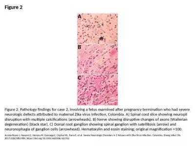
/
LK Cogs 107A Study materials for exam 1  1042006   Location termin LK Cogs 107A Study materials for exam 1  1042006   Location termin

LK Cogs 107A Study materials for exam 1 1042006 Location termin - PDF document

isabella
isabella . @isabella
Follow
342 views
Uploaded On 2022-08-19

LK Cogs 107A Study materials for exam 1 1042006 Location termin - PPT Presentation

rachnoid ura Mater The meninges were discussed in lecture Make sure you know the information which you were given Brain stem The brainstem is located at the juncture of the cerebrum and the spin ID: 938177

function location motor brain location function brain motor materials cogs located 107a study exam 2006 functions superior area cortex

Share:

Link:

Embed:

Download Presentation from below link

Download Pdf The PPT/PDF document "LK Cogs 107A Study materials for exam 1 ..." is the property of its rightful owner. Permission is granted to download and print the materials on this web site for personal, non-commercial use only, and to display it on your personal computer provided you do not modify the materials and that you retain all copyright notices contained in the materials. By downloading content from our website, you accept the terms of this agreement.


Presentation Transcript

(LK) Cogs 107A Study materials for exam 1 10/4/2006 Location terminology (rostral, caudal, anterior, posterior, lateral, medial, ventral, dorsal) Section definitions (midsagittal, parasagittal, coronal, horizontal) Lobes (frontal, temporal, parietal, occipital, limbic), regions in each, and dividing sulci (longitudinal fissure, lateral (Sylvian) fissure, calcarine sulcus, central sulcus, parietooccipital sulcus) Major divisions of CNS: myelencephalon through telencephalon The meningeal layers and CSF – see lecture materials Location of four main ventricles, choroid plexus – see lecture materials Must know location (what major division of the brain they’re in AND be able to locate on drawing of brain) and function of the following: medulla, pons, cerebellum, inf. and sup. colliculi, hippocampus, thalamus, hypothalamus, amygdala, corpus callosum, limbic cortex, basal ganglia, M1, S1, V1, A1, IT, Broca’s area, Wernicke’s area. The meninges “PAD” the brain. rachnoid,

ura Mater The meninges were discussed in lecture. Make sure you know the information which you were given. Brain stem: The brainstem is located at the juncture of the cerebrum and the spinal column. It consists of the midbrain , medulla oblongata , and the pons Limbic cortex, Components of limbic system - – see lecture materials (LK) Cogs 107A Study materials for exam 1 10/4/2006 The information below is a little more detailed than you need to know. But, make sure you know the structures which make up each brain division, the location of each, and a short summary of the function of each structure which is included in the list above (plus its location). Location: The prosencephalon is the most anterior portion of the brain. Structures:The prosencephalon consists of the telencephalon (cerebral cortex, basal ganglia), diencephalon (thalamus , hypothalamus , epithalamus, the optic tracts, optic chiasma, infundibulum, Ventricle III, mammillary bodies, posterio

r pituitary gland ), lateral ventricle and third ventricle Epithalamus: The is a dorsal posterior segment of the diencephalon (a segment in the middle of the brain also containing the hypothalamus and the thalamus ) which includes the habenula the stria medullaris and the pineal body . Its function is the connection between the limbic system other parts of the brain. Some functions of its components include the secretion of melatonin by the pineal gland, and the regulation of hunger and thirst by the habenula. Function:Controls Responses to Sight Eye Movement Pupil Dilation Body Movement Hearing Location: The mesencephalon is the most rostral portion of the brainstem . It is located between the forebrain and brainstem. Structures: The mesencephalon consists of the tectum (superior (visual) and inferior (auditory) colliculi) and tegmentum (cerebral aqueduct, periaqueductal gray, reticular formation , substantia nigra and the red nucleus). mi

dbrain Function:Autonomic Functions Complex Muscle Movement Conduction Pathway for Nerve Tracts Reflex Movement Simple Learning Location: The rhombencephalon is the inferior portion of the brainstem Structures: The rhombencephalon is comprised of the metencephalon , the myelencephalon , and the reticular formation (LK) Cogs 107A Study materials for exam 1 10/4/2006 MetencephalonFunction:Arousal Assists in Controlling Autonomic Functions Relays Sensory Information between the Cerebrum and Cerebellum Location: The pons is the portion of the brainstem that is superior to the medulla oblongata . The metencephalon is located below the posterior portion of the cerebrum and above the medulla oblongata Structures: The metencephalon is the division of the hindbrain that consists of the pons . Myelencephalon; Function: Controls autonomic functions Relays nerve signals between the brain and spinal cord Location: The medulla oblongata is the lower port

ion of the brainstem . It is inferior to the pons and anterior to the cerebellum Structures: The myelencephalon is composed of the medulla oblongata . medulla Function: Motor Control Receives Auditory, Somatosensory and Visual Sensory Signals Relays Sensory Signals to the Cerebral Cortex Location: The thalamus is a large, dual lobed mass of grey matter cells located at the top of the brainstem superior to the hypothalamus . The blue structure is the thalamus (LK) Cogs 107A Study materials for exam 1 10/4/2006 Hypothalamus Function:Controls Autonomic Functions Emotions Endocrine Functions Homeostasis Motor Functions Regulates Food and Water Intake Regulates Sleep Wake Cycle Location:The hypothalamus is located below the thalamus and posterior to the optic chiasma. The blue structure is the hypothalamus Involved in visual reflexes, and they receive input from: the eyes; the inferior colliculi; the skin; and the cerebrum. The Superior Collicul

i regulate the reflexive movement of the eyes and head, in response to a number of different stimuli. Fibers from the Superior Colliculi project to cranial nerve nuclei and to the superior cervical portion of the spinal cord where they stimulate motor neurons involved in turning the eyes (Oculomotor, Trochlear Abducens Cranial Nerves) and the head (the accessory cranial nerve and superior cervical cord levels). Impulses reaching the Superior Colliculi from the Cerebrum are involved in the visual tracking of moving objects. Involved in hearing and are an integral portion of the auditory pathways in the CNS. Neurons conducting impulses from the structures of the inner ear to the brain, all synapse in the Inferior Colliculi. Function: translates plans from M1 into specific instructions, timing, helps control saccades, compares current motor plan to new plans sent from other brain areas. (straight from Cogs 107A lecture – know this!) Location: Located just above th

e brainstem , beneath the occipital lobes at the base of the skull. cerebellum (LK) Cogs 107A Study materials for exam 1 10/4/2006 Amygdala Function:Arousal Controls Autonomic Responses Associated with Fear Emotional Responses Hormonal Secretions Location: The amygdala is an almond shaped mass of nuclei located deep within the temporal lobes medial to the hypothalamus and adjacent to the hippocampus . The round red object is the amygdale Function:Consolidation of New Memories Emotions Spatial Orientation Location: The hippocampus is a horseshoe shaped sheet of neurons located within the temporal lobes adjacent to the amygdala . Note: the “hippocampal formation” includes the hippocampus and some other structures. The purple structure is the hippocampus (the red structure is the amygdale) Includes caudate, putamen, globus pallidus, substantia nigra, subthalamic nucleusperform many functions like skilled movements (learned motor sequences), posture,

may coordinate different regions of the brain, control muscle force and movement intensivty, and regulation of emotion and reward via dopamine (straight from Cogs 107A lecture – know this!) Other groupings you may hear are the striatum (caudate + putamen), the corpus striatum (striatum + globus pallidus), or the lenticular nucleus (putamen + globus pallidus) Location:The basal ganglia is located deep within the cerebral hemispheres in the telencephalon region of the brain. (LK) Cogs 107A Study materials for exam 1 10/4/2006 Function:Controls Facial Neurons Involved in speech production and processing language Location: Broca's area is located in the left frontal lobe, around the opercular and triangular sections of the inferior frontal gyrus Function: Language Comprehension (note: this is a very very simplified summary of function!) Location: Wernicke's area is located in the left temporal lobe , posterior to the primary auditory complex. Corpus call

osum Function: Connects the left and right Hemispheres Location: The corpus callosum is a thick band of fibers located between the cerebral hemispheres. The pink structure is the corpus callosum (medial view) Very BRIEF summary of functions of some of the areas mentioned above SectionFunction Spinal Cord Input-output of sensory and motor information to and from the CNS-PNS Medulla Autonomic function (breathing, heart rate, etc.) Pons Auditory and vestibular (balance), sensory and motor Cerebellum Motor coordination and motor learning Midbrain Visuomotor functions, visual reflexes, auditory relays, motor coordination Thalamus Part of the diencephalon within the forebrain. Projects information to specific areas of the cerebrum, and controls which information is sent to the cerebral cortex Hypothalamus Regulates homeostasis in conjunction with the autonomic nervous system Basal Ganglia Centers for motor coordination Hippocampus Memory formation Amygdala E

motion processing Corpus Collosum Bundle of axons which connects the two hemispheres (LK) Cogs 107A Study materials for exam 1 10/4/2006 Limbic Lobe (outlined in white) (LK) Cogs 107A Study materials for exam 1 10/4/2006 The location of primary sensory and association areas of the human cerebral cortex. The primary auditory cortex is mostly hidden from view within the Sylvian fissure. From Guyton (1987). Note: primary motor cortex is in the gyrus just anterior to the central sulcus. (LK) Cogs 107A Study materials for exam 1 10/4/2006 A depiction of the left hemisphere of the brain showing the main language areas. The area in the inferior frontal lobe is known as Broca’s area, and the area in the superior temporal lobe is known as Wernicke’s area, named after the 19th century physicians who first described their roles in language. Broca’s area is adjacent to the motor cortex and is involved in (LK) Cogs 107A Study materials for exam 1 10/4