/
IMMUNOLOGY P IMMUNOLOGY P

IMMUNOLOGY P - PDF document

ivy
ivy . @ivy
Follow
343 views
Uploaded On 2022-08-27

IMMUNOLOGY P - PPT Presentation

RACTICAL week 10 
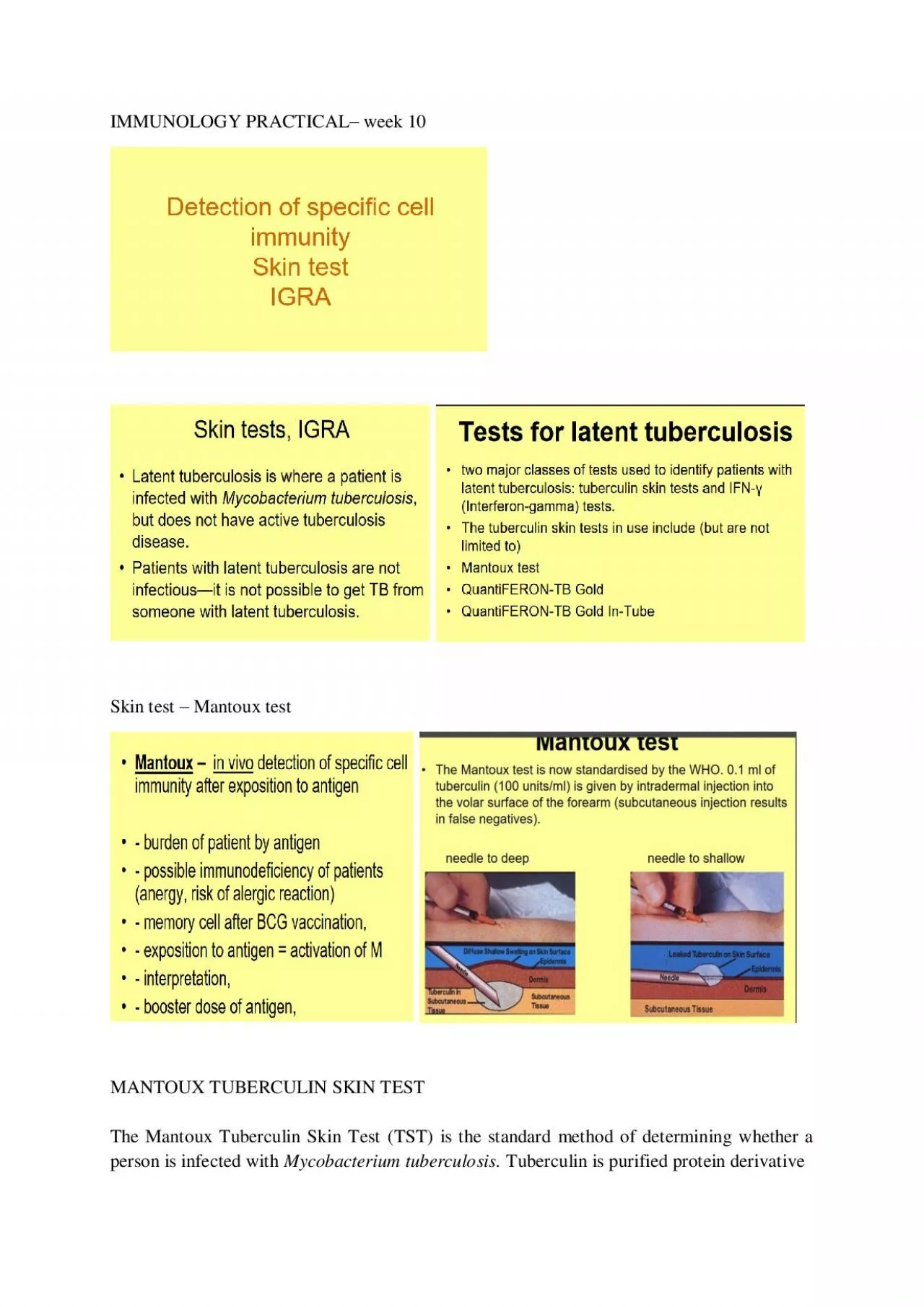
Skin test Mantoux test MANTOUX TUBERCULIN SKIN TEST The Mantoux Tuberculin Skin Test TST is the standard method of determining whether a person is infected with Mycobac ID: 942443

skin test infection tuberculin test skin tuberculin infection tuberculosis tuberculosi reaction tests infected ppd tst blood disease release bcg

Share:

Link:

Embed:

Download Presentation from below link

Download Pdf The PPT/PDF document "IMMUNOLOGY P" is the property of its rightful owner. Permission is granted to download and print the materials on this web site for personal, non-commercial use only, and to display it on your personal computer provided you do not modify the materials and that you retain all copyright notices contained in the materials. By downloading content from our website, you accept the terms of this agreement.


Presentation Transcript

IMMUNOLOGY P RACTICAL – week 10 Skin test – Mantoux test MANTOUX TUBERCULIN SKIN TEST The Mantoux Tuberculin Skin Test (TST) is the standard method of determining whether a person is infected with Mycobacterium tuberculosis. Tuberculin is purified protein derivative (PPD), an extract of Mycobacterium tuberculosis, M. bovis , or M. avium that is used in skin testing in animals and humans to identify a tuberculosis infection. PPD is a poorly defined, complex mixture of antigens.Tests based upon PPD are relatively unspecific since many of its proteins are fo und in different mycobacterial species. The tuberculin skin test is based on the fact that infection with M. tuberculosis bacterium produces a delayed - type hypersensitivity skin reaction. The components of the organism are contained in extracts of culture f iltrates and are the core elements of the classic tuberculin PPD, that is used for skin testing for tuberculosis. Reaction in the skin to tuberculin PPD begins when T - cells, whi

ch have been sensitized by prior infection, are recruited to the skin site wher e they release lymphokines. These lymphokines induce induration (a hard, raised area with clearly defined margins at and around the injection site) through local vasodilation leading to fluid deposition known as edema, fibrin deposition, and recruitment of other types of inflammatory cells to the area. Tuberculin Skin Test – Administration The TST is performed by injecting 0.1 ml of tuberculin - purified protein derivative (PPD) into the inner surface of the forearm. The injection should be made with a tuberculi n syringe, with the needle bevel facing upward. The TST is an intradermal injection. When placed correctly, the injection should produce a pale elevation of the skin (a wheal) 6 to 10 mm in diameter. Tuberculin Skin Test - Reading The skin test reaction shoul d be read between 48 and 72 hours after administration. A patient who does not return within 72 hours will need to be rescheduled for another skin test. The reaction sho

uld be measured in millimeters of the induration (palpable, raised, hardened area or sw elling, .The reader should not measure erythema (redness). The diameter of the indurated area should be measured across the forearm (perpendicular to the long axis). Tuberculin Skin Test - Interpretation Skin test interpretation depends onthe measure ment in millimeters (mm) of the induration andthe person’s risk of being infected with TB and/or progression to disease if infected. The following three cut points should be used to determine whether the skin test reaction is positive. A measur ement of 0 mm or anything below the defined cut point for each category is considered negative. Tuberculin Skin Test - False - positive ReactionSome persons may react to the TST even though they are not infected with M. tuberculosis. The causes of these false - p ositive reactions may include, but are not limited to, the following: Infection with nontuberculosis mycobacteria, previous BCG vaccination, incorrect method of TST administration, incorrect

interpretation of reaction and incorrect bottle of antigen used. INTERFERO N - GAMMA RELEASE ASSA Y (IGRA ) Interfero n - Gamma Release Assays (IGRAs) are whol e - blood tests that can aid in diagnosing Mycobacterium tuberculosi s infection. They do not help differentiate latent tuberculosis infection (LTBI) from tuberculosis dise ase. Two IGRAs are commercially availa b l e : • QuantiFERON ® - TB Gold I n - Tube test (QF T - GIT ) • T - SPOT® . T B test ( T - Spot ) IGRAs measure a person’s immune reactivity to M. tuberculosi s . White blood cells from most persons that have been infected with M. tuberculosi s wil l release interfero n - gamma (IF N - γ ) w hen mixed with antigens (substances that can produce an immune response) derived from M. tuberculosi s . To conduct the tests, fresh blood samples are mixed with antigens and controls. Positive IGRA : This means that the per son has been infected with M. tuberculosis . Additional tests are needed to det

ermine if the person has latent TB infection or TB disease. A health care worker wi l l then provide treatment as needed . Negative IGRA : This means that the person’s blood did not re act to the test and that latent TB infection or TB disease is not likely . IGRAs are the preferred method of TB infection testing for the following : • People who have received bacille Calmett e – Guérin (BCG ) . (BCG is a vaccine for TB disease) . • People who have a difficult time re t urning for a second appointment to look for a reaction to the TST . QuantiFERON ® - TB Gold I n - Tube tes t The QuantiFERON ® - TB Gold I n - Tube (QF T - G) is a blood test for use as an aid in diagnosing Mycobacterium tuberculosi s infection (both latent t uberculosis in f ection and active tuberculosis disease) . The QF T - G is an indirect test for M. tuberculosi s infection that is based on measurement of a cel l - mediate d immune response. A cocktail of 3 mycobacterial proteins ( 2 6 ESA T - 6, 2 7 CF P - 10, and

TB 7 , 7) sti mulate the patient's T - cells in vitr o to release interfero n - gamma, which is then measured using ELISA technology. The test detects infections produced by the M. tuberculosi s complex (including M. tuberculosi s , M. bovi s , and M. africanu m infections). BCG str ains and the majority of other no n - tuberculosis mycobacteria do not harbor ESA T - 6, CF P - 10, and TB 7 ,7 proteins , thus, patients either vaccinated with BCG or in f ected with most environmental mycobacteria should test negative. Results should always be interpr eted in conjunction with other clinical and laboratory findings . Sources: Kompani ková, Neuschlová, Sadloňová : Special B acteriology – Basic Laboratory T ests Available from: https://www.jfmed.uniba.sk/fileadmin/jlf/Pracov iska/ustav - mikrobiologie - a - imunologie/VLa/Special_Bacteriology_ - _basic_laboratory_tests.pdf https://www.jfmed.uniba.sk/fileadmin/jlf/Pracoviska/ustav - mikrobiologie - a - imunologie/VLa/imu_pract_7_skin_tests__IGRA.p