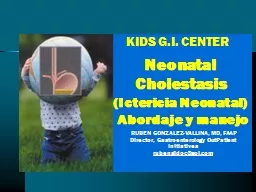
Director Gastroenterology OutPatient Initiatives rubengidocaolcom Neonatal Cholestasis Ictericia Neonatal Abordaje y manejo BREAST MILK JAUNDICE Early Onset Very common 12 percent of breastfed babies affected ID: 913877
Download Presentation The PPT/PDF document "KIDS G.I. CENTER RUBEN GONZALEZ-VA..." is the property of its rightful owner. Permission is granted to download and print the materials on this web site for personal, non-commercial use only, and to display it on your personal computer provided you do not modify the materials and that you retain all copyright notices contained in the materials. By downloading content from our website, you accept the terms of this agreement.
Slide1
KIDS
G.I. CENTER
RUBEN GONZALEZ-VALLINA, MD, FAAPDirector, Gastroenterology OutPatient Initiativesrubengidoc@aol.com
Neonatal Cholestasis(Ictericia Neonatal) Abordaje y manejo
Slide2BREAST MILK JAUNDICE
Early Onset
Very common – 12 percent of breast-fed babies affected.Onset during the first three days of life.May be related to caloric deprivation and dehydration.
Slide3Late Onset
Occurs in approximately 0.5 to 3 percent of healthy newborn.Serum bilirubin rises rapidly after the fourth day of life.Serum bilirubin peaks at the end of the second week of life.Severe jaundice (bilirubin
15 mg per deciliter) occurs in 2 percent of breast-fed babies and 0.3 percent of formula-fed babies. BREAST MILK JAUNDICE
Slide4Not associated with
kernicterus.Cessation of breast-feeding for 24 to 48 hours results in a significant drop in serum bilirubin.BREAST MILK JAUNDICE
etiology [?]abnormal progesterone metabolite
Inhibition of conjugating mechanism↑ levels of nonesterfied FA
↑ absorption of bile from the GI tract
Slide5JAUNDICE – BREAST FEEDING
Bilirubin may to 20mg/dl = in term infantsNeed to interrupt feeding = extremely rareResolves spontaneously = without interruptionIf bilirubin is too high = interrupt for up to 3 days / bilirubin will Minimal rebound
= after reinstitution of breast feeding
Slide6A breast-fed infant is found to have an
indirect bilirubinlevel of 15 mg/dL on day 15 of life. Of the following, thestatement which most correctly describes hyperbilirubinemia
as aresult of ingestion of breast milk is: (A) Excess bilirubin production is the primary etiologic factor. (B) A bilirubin level >10 mg-dL on the first day of life is typical
(C) The condition will resolve spontaneously (D) Affected infants are at risk of kernicterus (E) The bilirubin is transmitted in the breast milk.
Slide7Early Signs:
Poor suckingLethargy Late Signs:SeizuresFeverDirect bilirubin does not give kernicterusKERNICTERUS
Slide8JAUNDICE
BREAST FEEDINGVITAMIN K DEFICIENCYVitamin K Content
Commercial formulas = 50 mcg/LHuman milk = <1mcg/L
Slide9JAUNDICE – BREAST FEEDING
HEMORRHAGIC DISEASE -NEWBORNClassification
Classic = 2-5 days of life infant=not receive prophylaxis of Vit K Late= 2-12 wks of age
Slide10Prolongation of
2nd to (II, VII, IX, X)
In contrast congenital defects: PTT PT normalDIC (disseminated intravascular coagulation): platelets fibrinogen BREAST FEEDING VITAMIN K DEFICIENCY
PT
PTT
In the absence of vitamin K, these factors do not bind Ca
2+ normally and their coagulation activity is greatly reduced
Slide11Breast Feeding Vitamin K Deficiency
Sample Questions1 month born at home = with subdural hematoma PT, PTT =
Platelets, fibrinogen = normal3 day old born at home = bloody stools6 week old born at home = diarrhea bloody stools skin bruises
Deficiency of Factors II, VII, IX, and X
Slide12CONJUGATED
HYPERBILIRUBINEMIAALWAYS: PATHOLOGIC IN NEONATESEVALUATION
: MANDATORYGOALS:IDENTIFYINFECTIOUSMETABOLICCONGENITALGENETICOBSTRUCTION
Slide13DIFFERENTIAL DIAGNOSIS
OFCONJUGATED HYPERBILIRUBINEMIA
Extrahepatic ObstructionInfantile Obstructive CholangiopathyBiliary atresiaNeonatal hepatitisCholedochal cystOther causesBile plug syndromeCholedocholithiasis
Spontaneous bile duct perforationExtrinsic bile duct compressionGenetic and Metabolic DisordersDisorders of carbohydrate metabolismGalactosemiaFructosemiaGlycogen storage disease type IVDisorders of amino acid
metabolism
Tyrosinemia
Disorders of lipid metabolism
Niemann
-Pick disease
Gaucher
disease
Wolman disease
Cholesterol ester storage disease Chromosomal disordersTrisomy 18Down syndromeMiscellaneous genetic & metabolic disorders α1-Antitrypsin deficiencyNeonatal hypopituitarismCystic fibrosisZellweger cerebrohepatorenal syndrome
Familial hepatosteatosis
Persistent Intrahepatic Cholestasis
Paucity of intrahepatic bile ductsArteriohepatic
dysplasiaBenign recurrent intrahepatic cholestasisByler diseaseHereditary cholestasis with lymphedema
Trihydroxycoprostanic
academia
Acquired Intrahepatic Cholestasis
Infections
Hepatitis B (non-A, non-B?)
Syphilis
Toxoplasmosis
Rubella
Cytomegalovirus
Herpes
Varicella
Echovirus
Coxsackievirus
Leptospirosis
Tuberculosis
Bacterial sepsis
Drug-induced cholestasis
Cholestasis associated with parenteral nutrition
Slide14Slide15Neonatal
CHOLESTASISPhysiologic definition: Decreased bile flowIncreased in premature infants Cholestatic
Jaundice indicates hepatobiliary dysfunction.Conjugated hyperbilirubinemia developing within the first 90 days of life.Conjugated bilirubin > 1.0 mg/dl Incidence: 1 in 2500 live births US
Slide16Slide17IDIOPATHIC NEONATAL
HEPATITISMOST COMMON DX: IN CHOLESTATIC NEONATES
ETIOLOGY: UNKNOWNDX: OF EXCLUSIONCLASSIFICATION:SPORADICFAMILIAL (10%
POOR PROGNOSIS) ?HEREDITARY VS. METABOLIC FACTORSINCIDENCE (1:4800/ 1:9000) LIVE BIRTHS
Slide18NEONATAL HEPATITIS
CLINICAL PRESENTATIONS↑ MALESLOW BIRTH WEIGHT
50% = JAUNDICE 1ST WEEK OF LIFELFT’S: ↑ MILD TO MODERATE (2-10x
)1/3 = FTT, MORE COMPLICATED COURSE, LOOKS ILL, DECREASED APPETITE, AND DECREASED
ACTIVITY
ACHOLIC STOOLS
=
NOT COMMON
LIVER
:
ENLARGED
FIRM
Slide19TREATMENT
CONSERVATIVEPROGNOSIS :
GOOD
Slide20NEONATAL HEPATITIS
SAMPLE QUESTION4 week old maleMild
jaundice since birthPoor weight gainPale yellow – green color stoolsNext step: fractionate bilirubin (direct or indirect)DX: Neonatal HepatitisRSV is not associated with Neonatal HepatitisTorch
Titers should be done
Slide21BILIARY ATRESIA
1/3 OF CASES OF NEONATAL CHOLESTASISINCIDENCE:
1: 15000 (400-500 INFANTS PER YEAR)FEMALES: (4 x) MORE FREQUENT
FAMILIAL INCIDENCE: RARE
Slide22BILIARY ATRESIA
IS A DYNAMIC AND PROGRESSIVE
PANDUCTULAR SCLEROTIC PROCESSETIOLOGY UNKNOWN REOVIRUS TYPE 3 (?)
Slide23TYPES OF
BILIARY ATRESIA
Slide24BILIARY ATRESIA
CLINICAL PRESENTATIONSHINT
OBSTRUCTIVE JAUNDICEPATIENT LOOKS WELLPARENCHYMAL OR METABOLIC DISEASEPATIENT LOOKS ILL
Slide25BILIARY ATRESIA
CLINICAL PRESENTATIONSJAUNDICE: 3-5 WEEKS AFTER BIRTH
LFT’S: MILD ELEVATION UP TO 5x (USUALLY IN THE 200 RANGE, 150-300).BILIRUBIN: 14-15 RANGE, MOSTLY DIRECT
ALK PHOSPHATASE: MARKEDLY ELEVATEDHEPATOSPLENOMEGALYACHOLIC STOOLS: COMMON
Slide26Slide27BILIARY ATRESIA
ASSOCIATED ANOMALIES(10-15%)PREDUODENAL PORTAL VEIN
MALROTATIONPOLYSPLENISM OR HYPOSPLENISMABSENT INFERIOR VENA CAVABILOBED RT LUNGTEFESOPHAGEAL ATRESIA
Slide28EVALUATION OF THE INFANT WITH CHOLESTASIS
Slide29Slide30INTRAOPERATIVE CHOLANGIOGRAM
Intraoperative cholangiogram shows no evidence of cystic duct or gallbladder. No common bile duct stone is present.
Slide31BILIARY
ATRESIA PROGNOSISUNTREATED < 2 YEARS
KASAI: 33% 10 YR SURVIVAL RATE75% < 2 MONTHS10%
>3 MONTHS
Slide32Slide33Slide34BILIARY ATRESIA
SAMPLE QUESTIONS4 week old = female, male
Looks well except that appear yellowMild hepatosplenomegalyTB = 10, LFTS = 200 rangeNext Step: URGENT to differentiate between
intra/extrahepatic obstruction and fractionate bilirubin (direct or indirect)
Slide35BILIARY ATRESIA
SAMPLE QUESTIONSFT female infant
5th day of life jaundiceAppears healthy otherwiseThe best study to evaluate bile excretion = PIPIDA (hida) = no excretion
Next step = liver biopsy
Slide36ALPHA1-ANTITRYPSIN
IS A GLYCOPROTEINPRODUCED BY HEPATOCYTES (2 GRAMS PER DAY)SERINE – PROTEINASE INHIBITOR
INHIBITOR OF: NEUTROPHIL ELASTASE ACID PROTEASES OF ALVEOLAR MACROPHAGES
Slide37Slide38ALPHA 1 ANTITRYPSIN DEFICIENCY
Pi PhenotypeMMZZNullMmalton
NduharteMZSZMSRiskO10-20%0?
RISK FOR LIVER DISEASE
25%
= LIVER DISEASE
25% = LUNG DISEASE25%
= LIVER AND LUNG DISEASE
25%
= NO DISEASE
Slide39ALPHA1-ANTITRYPSIN
INCIDENCEARMOST COMMON GENETIC CAUSE OF LIVER
DISEASE IN CHILDRENCOMMON IN WHITESNORTHERN EUROPEAN AND SCANDINAVIANIN THE U.S. 1/2000
IS HOMOZYGOUS 1/30 IS HETEROZYGOUS
Slide40ALPHA1-ANTITRYPSIN
CLINICAL FEATURES10% OF PATIENTS ZZLACK OF SPECIFIC CLINICAL FEATURES
IN NEONATES: PROBABLY DAMAGE BEGAN IN UTERUS45% SGACOMMON: POOR FEEDING, IRRITABILITY, FIT LETHARGYJAUNDICE = 3-12 WEEKS
RESOLUTION IN 50% BY 6-8 MONTHSREMEMBER, THE PATIENT IN METABOLIC DISEASE OR PARENCHYMAL DISEASE LOOKS SICK.
Slide41ALPHA1-ANTITRYPSIN
PHYSICAL EXAMINATIONNOT SPECIFICPATIENT UNDERNOURISHED
HEPATOMEGALYSMOOTHFIRMSPLENOMEGALYFREQUENT BRONCHITIS OR PNEUMONIA
REMEMBER TO ASK IF SOMEONE IN THE FAMILY HAS DIED OF EMPHYSEMA AT A YOUNG AGE
Slide4225% = LIVER DISEASE
25% = LUNG DISEASE25% = LIVER AND LUNG DISEASE25% = NO DISEASE
Slide43ALPHA 1
ANTITRYPSIN DEFICIENCYSAMPLE QUESTIONS
Alpha 1-antitrypsin level and phenotypeAlways remember to get a phenotype because level could be normal, or in infections. It is an acute phase reactant.
10 year old with no PMH; presents with UGI bleedingHepatosplenomegalyAscites plateletsMild in
LFTs, GGTP
PT
Test of diagnosis?
Slide449 month old with
FH = + emphysema
Hepatomegaly+ icterusLooks illTest for diagnosis?Alpha 1-antitrypsin level and phenotypeAlways remember to get a phenotype because level could be normal in infections.
It is an acute phase reactant. ALPHA 1ANTITRYPSIN DEFICIENCYSAMPLE QUESTIONS
Appetite
Irritability
lethargy
Slide45ALPHA
1ANTITRYPSIN DEFICIENCYSAMPLE QUESTIONS
serum alpha 1 antitrypsin level is associated with early cirrhosisMost common manifestation in infancy
Hepatic cirrhosis
Slide46GALACTOSEMIA
Galactose-1-phosphate uridyl transferase
= deficiencyIncidence >1 : 40,000GalacoseGalactose-1-phosphate accumulates in
Galactitol Lactose G
lucose and Galactose
Galactose
_galactose-1-phosphate
uridyl
transferase__
G
lucose
LiverKidneyEyesCNS
Slide47GALACTOSEMIA
SIGNS & SYMPTOMSAnorexiaLethargy
VomitingPoor Weight GainJaundiceHepatomegaly
CataractsAscitiesEarly CirrhosisRisk of sepsis If patient develops Group B Strep sepsis, it is galactosemia until proven otherwise
Symptoms begin within 2 weeks after ingestion of lactose containing
formulas.
Slide48GALACTOSEMIA - LABS
Mild cirrhosisHypoglycemiaMixed hyperbilirubinemia
LFTs PTRenal FanconiGalactose detected in urine as a non-reducing sugar
AcidosisGlycosuriaProteinuria Aminoaciduria
Slide49GALACTOSEMIA DIAGNOSIS
Measure in RBC’s = galactose-1- phosphate uridyl activityTreatment =
remove galactose from diet
Slide50GALACTOSEMIA
SAMPLE QUESTIONS2 week old breast fed
appetiteJaundice, hepatomegalyGlucose oxidate test in urine = (-)Check reducing substance in urine, order a C
linitestGalactose detected in urine as a non-reducing sugar
Slide51GALACTOSEMIA
SAMPLE QUESTIONS4 week old V + D; multiple cow’s
milk formula changes AppetiteLethargicJaundice, hepatomegalyOrder:Galactose-1-phosphate uridyl transferase activity in RBCs
Diagnosis:GalactosemiaTreatment:Soy formula
Slide52GALACTOSEMIA
SAMPLE QUESTIONS2-3 month breast fed; prolonged jaundiceV + D
Hepatomegaly, HypotoniaInitial test = check reducing substanceOrder a galactose-1-phosphate uridyl transferase activity in RBCs
Slide53HEREDITARY FRUCTOSE
INTOLERANCEIncidence: 1:20,000ARDeficiency:
Adolase BLiverKidneySmall BowelFailure to Split Fructose 1 Phosphate
Slide54SYMPTOMS
Expose to Fructose and SucroseDepends: Age
Fructose loadHINT: Fruits Juices Vegetables
Sucrose
Slide55SYMPTOMS
AcuteChronicNausea
& vomitingFailure to thriveTremorJaundice-cirrhosisDizzinessVomiting and diarrheaLethargy comaFeeding difficulties
Slide56DIAGNOSIS
Liver Bx: Fructose 1 Phosphate Aldolase
+ Reducing substance in the urineRemember: Look in the questions if the patient is on fruit juices? FRUCTOSE INTOLERANCE
Slide57Slide58THANK YOU
!!!!!!
Slide59REYES SYNDROME
UncommonIncidence: 3.5 / 100,000After viral infection
EmesisNon-icteric hepatic dysfunction
Slide60REYES SYNDROME
LABORATORIES ABNORMALITIES LFTs
(3-30 X normal) PT GlucoseIctericia = very rare NH
3 = 2-20 X normalIf >5 X normal = poor prognosis
Slide61REYES SYNDROME
Diagnosis = Liver BiopsyMicrovesicular steatosis
Mitochondrial alterations
Slide62REYES SYNDROME TREATMENT
Monitor glucose level ICP
Death usually from encephalopathy
Slide63Which
best treatment efforts improve prognosis? ICP
6-8 year old, vomiting, lethargic, responds to command LFT’s = 100 NH3 = 150 mg/ml
PT = 2-3 secComa stage IREYES SYNDROMESAMPLE QUESTIONSHINT:
Matching? Look
where
the glucose is the
lowest
and
bilirubin
is almost
normal
.
Slide64POISONING
CCL4
Causes Severe kidney and liver damageHINT: Matching? Look where the bilirubin and LFT’s are the highest
and High BUNWas used inExposure
Inhalation
Ingestion
Fire extinguisher
As a solvent
Anthelmintic
Slide65HEPATITIS A
RNA virus, transmitted fecal-orally (food +H
20)No transplacental transmissionNo carrier (occasionally prolonged cholestasis)Incubation is 15-50 daysAcute infection-high anti-HAV
IgMPrevious infection – high anti-HAV IgGSymptoms rare in children1% have a chance of fulminant hepatitis
IG
is good prophylaxis against HAV (
w/in 2 wks of exposure
Give IG
to
household and close
contacts
Slide66HEPATITIS A
SAMPLE QUESTIONSPre-icteric phase
Milder in children Anicteric infection may occurHINT: Biliary Atresia: Rare LFT’s are higher than 500Hepatitis A: LFT’s are usually higher 500
adults
anorexia
fever
Vomiting
Abdominal pain
Slide67HEPATITIS B
Hbs Ab = + = vaccinationHbs Ab = + Hbc Ab = +
Had Hepatitis B
Slide68HEPATITIS B
SAMPLE QUESTIONSInfant born to a mother HbsAg+
Treatment:Hep B immunoglobulin + vaccineBathed immediately after birthInfant risk = 70-90% in HbsAg+ mother 10-25% in HbsAg
- motherReduce vertical transmission by 95%
Slide69HEPATITIS B
SAMPLE QUESTIONSHepatitis Bs Ag = +No need to be vaccinated
Slide70CONSTIPATION
Majority is due to functional or behavioral problemSmall percentage
of children present with constipation have an organic causeSome breast-fed babies will stool 1/5-10 daysFunctional fecal retention is the most common nonorganic causeHINT: Rectal examination:
Rectal ampulla is full
Slide71HIRSCHPRUNG DISEASE
1/5000 birthsAbsence of enteric ganglionic neurons that begin at the anus and then extend proximallyIncreased association with
Down syndromeEvaluate any term infant that does not pass meconium within 48 hours of birthSuction rectal biopsy for diagnosis, full-thickness biopsy needed for unclear casesTreat with surgical resectionHINT: Rectal Ampulla is empty
KUB: No granular appearance or Ca2+ in ABD = CF yes
Slide72Typical Celiac Disease
Slide73Gastrointestinal Manifestations
(“Classic”)Most common age of presentation: 6-24 monthsChronic or recurrent diarrheaAbdominal distensionAnorexiaFailure to thrive or weight loss
Rarely: Celiac crisis Abdominal pain Vomiting Constipation Irritability
Slide74Non Gastrointestinal
ManifestationsDermatitis HerpetiformisDental enamel hypoplasia of permanent teethOsteopenia
/OsteoporosisShort StatureDelayed Puberty Iron-deficient anemia resistant to oral Fe Hepatitis Arthritis Epilepsy with occipital calcifications
Most common age of presentation: older child to adultListed in descending order of strength of evidence
Slide75Serological Test Comparison
Farrell RJ, and Kelly CP. Am J Gastroenterol 2001;96:3237-46. Sensitivity % Specificity %
AGA-IgG 69 – 85 73 – 90 AGA-IgA 75 – 90 82 – 95 EMA (IgA) 85 – 98 97 – 100 TTG (IgA) 90 – 98 94 – 97
Slide76HLA Tests
Potential role for DQ2/DQ8 asymptomatic relativesDown, Turner & Williams syndrometype 1 diabetesdiagnostic dilemmas
Slide77The
Antireflux barrier
Slide78Question..
Which of the following statements is true regarding reflux ?A. Thickening formula reduces reflux episodesB. Proton pump inhibitors have been found to improve infant irritabilityC. Treatment with PPI’s for three months is indicated in patients with
endoscopically proven reflux esophagitis D.Acute life threatening events have definitely been linked to gastroesophageal reflux diseaseE.Erythromycin has been proven to be beneficial in patients with GERD
Slide79Munchausen by P
roxy3 yo old female comes to the ER hx of severe constipation. The PE is normal. Mother demands an Xray
of the abdomen to be taken since that’s the way they dx constipation. Xray shows scattered stool around the colon. Mother insists on admission for clean out. The ER MD calls GI for advice for ? Go-Lytely clean out.A. The Abdominal Xray and the HPE are evidence she has constipation, needs a clean out.B. Parental insistence on admission may be reasonable given parents are knowledgeable.C. HPE and Xray do not support a dx of constipation. You will request a Hx of admissions, ER vistis and test results.
D. You recommend no admission: no indication to admit the patient. Refer the patient and mother to a psychiatrist.E. You recommend a CT scan of the abdomen since is more reliable than the Xray.
Slide80KIDS G.I. CENTER
RUBEN GONZALEZ-VALLINA, MD, FAAP