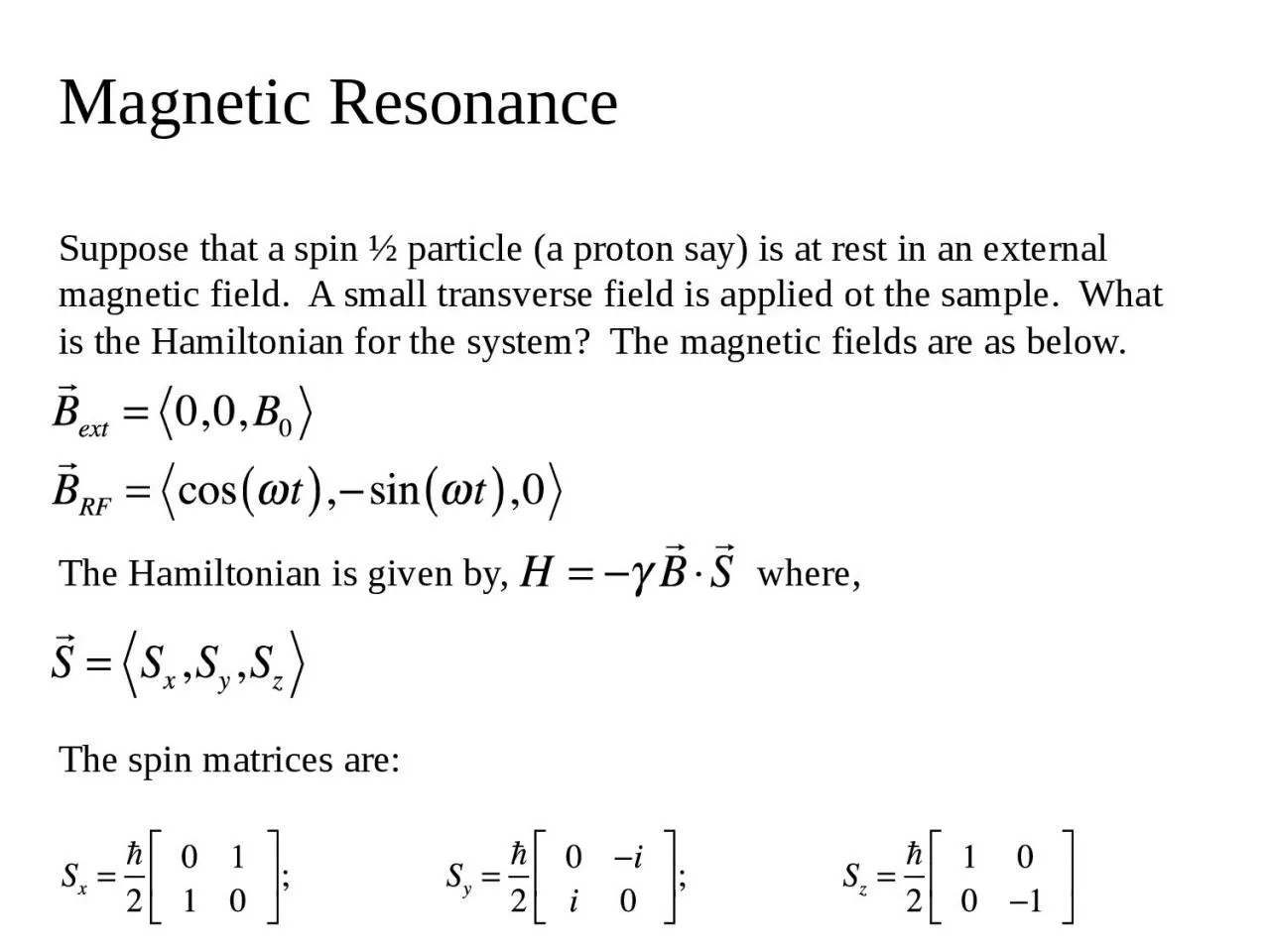
PPT-Magnetic Resonance Suppose that a spin ½ particle (a proton say) is at rest in an external

Author : jacey | Published Date : 2023-12-30

ot the sample What is the Hamiltonian for the system The magnetic fields are as below The Hamiltonian is given by where The spin matrices are Magnetic Resonance

Presentation Embed Code

Download Presentation

Download Presentation The PPT/PDF document "Magnetic Resonance Suppose that a spin ..." is the property of its rightful owner. Permission is granted to download and print the materials on this website for personal, non-commercial use only, and to display it on your personal computer provided you do not modify the materials and that you retain all copyright notices contained in the materials. By downloading content from our website, you accept the terms of this agreement.

Magnetic Resonance Suppose that a spin ½ particle (a proton say) is at rest in an external: Transcript


ot the sample What is the Hamiltonian for the system The magnetic fields are as below The Hamiltonian is given by where The spin matrices are Magnetic Resonance The Hamiltonian is. Principle first observed in 1946. Used for spectroscopy and imaging . Imaging techniques are a form of tomography, where slices are ’cut’ and depict. MRI utilizes signals from the body . MRI is non-ionizing, operating in radiofrequency range, unlike CT, PET, SPECT. Sarah Arveson. Magnetism. A charged particle will experience a force when placed in a magnetic field and given an initial velocity. This is given by the Lorenz force. where q is the charge of the particle in Coulombs, m is the mass of the particle in kilograms, v is velocity in meters per second, and B is the magnetic field in . 2.50 × 10. –28. kg. , and that of the other is . 1.67 × 10. –27. kg. If the lighter fragment has a speed of . 0.893. c. . after the breakup, what is the speed of the heavier fragment? . . An unstable particle at rest breaks into two fragments of unequal mass. The mass of the first fragment is . Department of Chemistry. Dylan W. Benningfield. Electron Spin Resonance (ESR). Electron spin resonance (ESR. ). Electron . paramagnetic resonance (EPR) . Study of paramagnetic materials. Radicals, bi-radicals, triplet states, unfilled conduction bands, transition metal ions, impurities in semi-conductors, etc.. 2.50 × 10. –28. kg. , and that of the other is . 1.67 × 10. –27. kg. If the lighter fragment has a speed of . 0.893. c. . after the breakup, what is the speed of the heavier fragment? . . An unstable particle at rest breaks into two fragments of unequal mass. The mass of the first fragment is . Part 1. Magnetic Resonance Imaging. A great multidisciplinary application of physics and other sciences. And example of a “pure science” discovery given a practical application. Based on NMR – Nuclear Magnetic Resonance – discovered in the 1940s. APS March Meeting, Mar 17, 2016. John S. Colton. Physics Department. Brigham Young University. Co-authors: . Kyle . Miller. . . Dr. Ross Spencer. Michael Meehan. Download the code at . http://www.physics.byu.edu/research/coltonlab/cavityresonance. Introduction to Spectroscopy. 2. Spectroscopy. is the study of the interaction of matter with the electromagnetic spectrum. Electromagnetic radiation displays the properties of both particles and waves. Electron can behave like a tiny bar magnet.. Atomic nucleus can spin and behave as a tiny bar magnet. These . spinning nuclei generate tiny magnetic . fields. Any nucleus will spin (odd mass or odd atomic number. Nuclear Magnetic Resonance Spectroscopy. Over the past fifty years nuclear magnetic resonance spectroscopy, commonly referred to as nmr, has become the preeminent technique for determining the structure of organic compounds. . 1. Introduction to Spectroscopy. 2. Spectroscopy. is the study of the interaction of matter with the electromagnetic spectrum. Electromagnetic radiation displays the properties of both particles and waves. ). B: Sagittal T2-weighted . TSE C. : Sagittal T2-weighted SE image . . Axial T2-weighted turbo spin-echo (TSE) (4902/132) magnetic resonance image of the uterus in the axial (A) and coronal (B) planes . ++. MRI(Magnetic Resonance Imaging). Radiation Safety (SEC). SUBJECT CODE – PHYS307TH. Magnetic Resonance Imaging (MRI). It is not possible to see the internal organs and structure of the human body directly to locate the infection or illness . Magnetic resonance imaging (MRI) is a spectroscopic imaging technique used in medical setting to produce image of the internal structure of the human body. The scan uses a strong magnetic field and radio waves to generate images of internal parts ( cells, tissues , organs ) of the body that can't be seen as well with X-rays , CT scans or ultrasounds . For example , it can help doctors to see inside joints , cartilage , ligaments , muscles and tendons , which make it helpful for detecting various sports injuries. It is also used to examine internal body structures and diagnose a variety of disorders , such as strokes, tumors , aneurysms , spinal cord injuries , multiple sclerosis and eye or inner ear problems. It is also widely used in research to measure brain structure and their function.. Spectroscopy. Learning Module #1. - The Electromagnetic Spectrum and Radiation. Learning Module #. 2. – Nuclear Magnetic . Resonace. (NMR) Spectroscopy . Learning Module #3. - Chemical Shift and Spin-Spin Multiplicity.

Download Document

Here is the link to download the presentation.
"Magnetic Resonance Suppose that a spin ½ particle (a proton say) is at rest in an external"The content belongs to its owner. You may download and print it for personal use, without modification, and keep all copyright notices. By downloading, you agree to these terms.

Related Documents