PDF-CARCINOMA OF THE APPENDIX
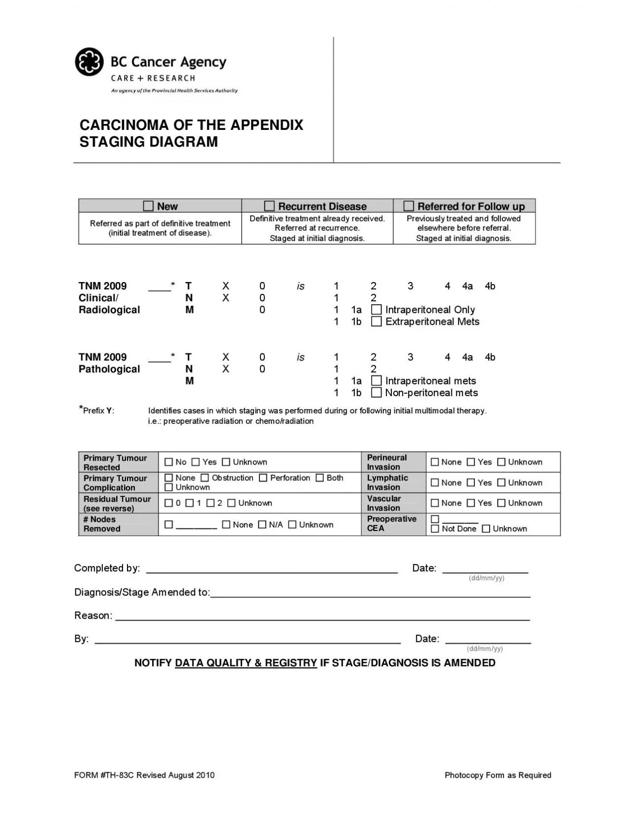
STAGING DIAGRAM New Recurrent Disease Referred for Follow up Referred as part of definitive treatment Definitive treatment already received Referred at recurrence
Download Presentation
"CARCINOMA OF THE APPENDIX" is the property of its rightful owner. Permission is granted to download and print materials on this website for personal, non-commercial use only, provided you retain all copyright notices. By downloading content from our website, you accept the terms of this agreement.
Presentation Transcript
Transcript not available.