/
h Intern Med Res 2021 4 2 142 h Intern Med Res 2021 4 2 142

h Intern Med Res 2021 4 2 142 - PDF document

jade
jade . @jade
Follow
343 views
Uploaded On 2022-08-22

h Intern Med Res 2021 4 2 142 - PPT Presentation

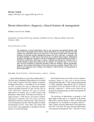
Ar c 14 8 DOI 1026502aimr00 6 5 Archives of Internal Medicine Research 143 Abstract Coronavirus disease COVID 19 was declared a pandemic in early 2020 Since then it has continued to spr ID: 940007

covid lesions 2020 chilblain lesions covid chilblain 2020 chilblains patients dermatol infection 2021 skin reported pernio pattern pandemic cases

Share:

Link:

Embed:

Download Presentation from below link

Download Pdf The PPT/PDF document "h Intern Med Res 2021 4 2 142" is the property of its rightful owner. Permission is granted to download and print the materials on this web site for personal, non-commercial use only, and to display it on your personal computer provided you do not modify the materials and that you retain all copyright notices contained in the materials. By downloading content from our website, you accept the terms of this agreement.


Presentation Transcript

Ar c h Intern Med Res 2021; 4 (2): 142 - 14 8 DOI: 10.26502/aimr.00 6 5 Archives of Internal Medicine Research 143 Abstract Coronavirus disease (COVID - 19) was declared a pandemic in early 2020. Since then, it has continued to spread havoc in the lives of millions of people. The Covid - 19 infection can have a spectrum of symptoms, ranging from asymptomatic infection to a fatal multi - organ failure. A number of unusual manifestations have also been reported. Among them are pernio - like lesions, also referred to as Chilblain - like lesions. Previously, th ese skin lesions were associated with damp conditions and autoimmune diseases. But, their presentation in this pandemic has confused the clinicians. It is very important to understand the clinical picture, pathophysiology, histology, and treatment of these lesions, especially in the COVID era. Hence, in this review, we have made an effort to understand these lesions further. The main aim of our article is to provide physicians with a detailed analysis of chilblains like lesions so that this can help them with diagnosis and treatment. Keywords: Chilblain - Like Lesions 1. Introduction The severe acute respiratory syndrome coronavirus two (SARS - CoV - 2), which causes the 2019 coronavirus disease (COVID - 19), has affected millions worldwide. It was declared a pandemic on 11 th march 2020 [ 1 ] and has not only affected the mental health of the general public but has also adversely affected the mental health of physicians involved in the care of these patients [2]. It typically presents with fever, cough, headache, diarrhea, myalgia, anosmia, asthenia, and sometimes pneumonia [3] . It can also lead to severe disease which results in acute respiratory distress syndrome (ARDS), renal failure, and a hyper - coagulable state which might end in stroke, myocardial infarction , or pulmonary embolism. However, few skin manifestations have also been described. These dermatological findings include maculopapular rash, urticaria, vesicular eruptions, chilblain - like lesions, and livedo/necrosis [4]. Chilblain - like lesions were the s econd most frequent cutaneous manifestation in the multicenter Italian study [5]. Chilblain - like lesions are also described as pernio - like lesions. These lesions initially present as erythematous - edematous or blistering skin lesions that mostly involve the extremities. Over the course of 1 - 2 weeks, these lesions become more purpuric and flattened, ultimately resolving without any treatment. The majority of COVID patients with chilblain - like lesions are mostly in good health, without any significant coron avirus symptoms, but they may have a history of upper respir

atory tract infection [6]. COVID - 19 - related chilblain - like lesions have been reported from Europe, Middle East, and the United States from February to May, but none have been reported from tropica l regions such as South America or Southeast Asia. A proposed theory is that cold exposure may be a co - factor for the cutaneous inflammation that occurs in chilblain - like lesions [4 ]. Type I interferon and cytokine response to the viral infection have be en shown to cause microangiopathic changes and chilblain - like lesions [7]. Due to potentially unreliable and subjective patient reporting, these lesions cannot be confirmed as a symptom of COVID - 19 disease. In some regions, due to limited coronavirus testi ng, the diagnosis of COVID - 19 and/or chilblain - like lesions may be missed or remain unreported. In this review, we have discussed in detail covid - 19 related pernio - like lesions. We have dig deeper and tried to summarize all the important details about this rare skin manifestation associated with the covid - 19 disease. 2. Methods and Results A review of the PubMed database was conducted on 14 Ar c h Intern Med Res 2021; 4 (2): 142 - 14 8 DOI: 10.26502/aimr.00 6 5 Archives of Internal Medicine Research 144 April 2021 using the search terms “Covid - 19,” “Pernio - like lesions, and “Covid - 19 chilblains”. Filter for humans and articles written in the English language was applied. The total number of articles retrieved was 154. These records were then screened by title and abstract content. Peer - reviewed publications that contained original patient cases of COVID - 19 and a discussion of the pernio - like lesions were included in the analysis. A total of 24 articles were included in our review. 3. Discussion Coronavirus is being considered the respiratory virus but othe r presentations are also possible, and some are frequently emerging [8]. COVID - 19 - related chilblain - like lesions were first reported in a 13 - year - old boy in early March by Italian authors [9]. Since then, several cases of chilblain - like acral lesions mostl y involving children and adolescents worldwide have been reported and published in the scientific literature [5]. Skin lesions have been classified as prodromal (before COVID - 19 onset), acute (within 2 weeks of COVID - 19 onset), post - acute (from week 2 to 4 ), or late (after week 4). Chilblain - like lesions mostly occur between weeks 2 and 4 [10] and have a mean duration of 12.7 days [11]. Ladha MA et al. summarized key findings in her article that the cold season is associated with COVID - 19 chilblains and that these lesions are asymmetrical and mainly affect the feet. Simulta

neous hand and feet involvement is very rare [12]. Rare cases of chilblain - like lesions involving other acral sites, such as the auricular region have also been reported as a patient presented with a single, red - purple, extremely painful, and infiltrated papule on the lateral side of the right auricle. She was tested positive for COVID - 19 infection 14 days ago but had an asymptomatic course [13]. Figure 1 and 2 show pernio like lesions as reported by Ladha MA et al [11] and Proietti I et al [13]. Figure 1: Clinical Photographs of Chilblain - like lesions [13]. Ar c h Intern Med Res 2021; 4 (2): 142 - 14 8 DOI: 10.26502/aimr.00 6 5 Archives of Internal Medicine Research 145 Figure 2: A rare case of auricle perniosis [14]. Few studies mentioned the occurrence of blistering lesions with these pernio - like lesions. Piccolo et al. reported the presence of blistering lesions in 23 out of 54 patients [14]. On the other hand, Colonna C et al. [15] and Freeman EE [16] did not find any such bullous lesions in their studies. Other commonly reported symptoms w ere pruritus, pain, and burning sensations [5 ]. Patients with COVID - 19 chilblains present with less severe COVID, with decreased rates of pneumonia, lower hospital and ICU admissions, and reduced mortality [17]. The majority of patients have complete resol ution of COVID - 19 chilblains without treatment and sequelae, though post inflammation hyperpigmentation may occur. Relapsing courses have also been reported [14]. Most patients do not have other systemic symptoms of COVID - 19 but, Piccolo et al. reported ga strointestinal symptoms (11%), respiratory symptoms (7%), and fever (4%) [17]. 3.1 Histopathological f indings Several studies have explained the histopathological features of chilblain - like lesions as observed in COVID infection. Navarro L et al. describes dermoscopy findings of 22 children with chilblains [18]. Interestingly, dermoscopy of these lesions revealed the presence of an indicative pattern represented by a red background area with purpuric globules and grey - brown reticul e. The study further elaborates that the red color in the background is related to the inflammatory infiltrate, hemosiderin deposition, and vascular dilatation, while globules represent extravasated red cells, and the grey - brown reticule is possibly due to pigment incontinence, lichenoid infiltrate along with vascular changes. On the other hand, S. Recalcati directly observed 14 cases including 11 children. Histology of the acral lesions showed a diffuse dense lymphoid infiltrate of the superficial and deep dermis and the hypodermis, with a prevalent perivascular patter

n, and signs of endothelial activation [19]. Anoth er study carried out by Sohier P et al. included 13 patients. Two main histopathologic patterns were observed: a chilblain - like histopathologic pattern (10 of 13 cases; 77%) and a thrombotic vasculopathy pattern (3 of 13 cases; 23%). The chilblain - like his topathologic pattern featured a superficial and deep perivascular infiltrate of lymphocytes of varying intensity. The thrombotic vasculopathy pattern featured an absent or mild inflammatory infiltrate, multiple intraluminal fibrin thrombi, and ischemic epi dermal necrosis [20]. Colmenero et al. took skin biopsies of 7 children with Ar c h Intern Med Res 2021; 4 (2): 142 - 14 8 DOI: 10.26502/aimr.00 6 5 Archives of Internal Medicine Research 146 chilblain - like acral lesions, performed immune - histochemistry and microscopic examination. He confirmed the presence of SARS - CoV - 2 in endothelial cells of blood vessels, suggestin g that virus - induced vascular damage and secondary ischemia could be the cause of these lesions [21]. On the other hand, Baeck M mentioned that even though positive anti - SARS - CoV/SARS - CoV - 2 immunostaining on skin biopsy of chilblains seems to confirm the p resence of the virus in the lesions, but it lacks specificity and must be interpreted with caution [22]. Table 1 summarizes all the above findings. Reference Subjects Procedure Findings Navarro L et al. [16] 22 Dermoscopy Lesions revealed the presence of an indicative pattern represented by a red background area with purpuric globules and grey - brown reticule. S Recalcati et al. [17] 14 Skin biopsy and histological examination Lesions showed a diffuse dense lymphoid infiltrate of the superficial and deep dermis, as well as hypodermis, with a prevalent perivascular pattern and signs of endothelial activation. Sohier P et al. [18] 13 Skin biopsy and histological examination A chilblain like histopathologic pattern and a thrombotic vasculopathy pattern was found. Colmenero et al. [20] 7 Immunohistochemistry and electron microscopy Virus - induced vascular damage and secondary ischemia. Table 1 : Histopathological findings . 3.2 Pathophysiological findings The exact pathogenesis of pernio - like lesions caused by COVID - 19 is unknown but endothelial damage, coagulation abnormalities and obliterative micro - angiopathy seem to be involved [23]. Recalcati S et al. mentioned that chilblain - like lesions associated with covid - 19 could be secondary to a delayed immune‐ mediated response involving the small cutaneous capillaries [19 ]. Increased interferon and cytokine - mediated inflammation due to COVID

- 19 could be another mechanism involved in the pathogenesis of these lesions. SARS‐CoV‐2 infection might induce the expression of type 1 interferon, leading to micro - angiopathic changes. Hubiche et al. demonstrat ed a significantly higher IFN - alpha response in patients with chilblain lesions [24]. Freeman EE et al. proposed that prothrombotic coagulopathy could be a contributing factor to these lesions, and it is well known that COVID - 19 infection can be complicate d by hypercoagulability and raised D - dimers and fibrinogen [16]. Furthermore, antiphospholipid antibodies have been observed in patients with acutely ill COVID - 19 infection, and interestingly these antibodies have previously been associated with pernio - lik e lesions [25]. 3.3 Treatments options Chilblain lesions have also been reported in association with other conditions such as viral hepatitis, malignancy, and autoimmune and hematologic diseases. They can also be caused by relatively benign conditions such as cold exposure. So previous research es mentioned treating the underlying cause for the treatment of chilblain lesions. Hot fomentation of the affected area is encouraged when the cause of the Ar c h Intern Med Res 2021; 4 (2): 142 - 14 8 DOI: 10.26502/aimr.00 6 5 Archives of Internal Medicine Research 147 chilblain is cold exposure. Other treatment options include corticosteroids and calcium channel bloc kers. Topical nitroglycerin use was also recommended by some studies [26]. There have been no studies to evaluate the treatment of these lesions in COVID - 19 patients. So, treatments for idiopathic and secondary chilblains can be extrapolated to COVID - 19 ch ilblains. Ladha MA et al. recommended potent topical corticosteroids and then escalating as required. Further recommendations according to the current understanding of the pathogenesis of these lesions include antiplatelet therapies such as low dose ASA, t o reduce clotting and distal ischemia and these can be considered in the absence of contraindications [12]. 4. Conclusion Pernio - like lesions are a common dermatological manifestation of COVID - 19. Mostly, these lesions are asymmetrical and limited to fee t but the involvement of other sites is also possible. They mostly occur in the post - acute phase of COVID - 19 infection. Although the link between COVID - 19 and these lesions is still uncertain, in the absence of cold - damp conditions and the presence of COVI D - 19 community spread, COVID - 19 should be considered as a cause of these lesions. By using this inference, we would be able to recognize asymptomatic covid - 19 patients, who are likely to spread infection within the community. Hence, all

patients with suspe cted COVID - 19 chilblains should follow public health guidelines for testing and self - quarantine. Furthermore, we suggest that larger population - based studies should be designed to fully understand the association between COVID - 19 and these pernio - like lesi ons. References 1. Wang CJ, Worswick S. Cutaneous manifestations of COVID - 19. Dermatol On line J 27 (2021):13030. 2. Anoshia Afzal, Maria Kamal, Neni Diyanti, e al . Implementation of Mandatory Counseling Sessions and Availability of Support Centers for Mental Well Being of Heath Care Workers during the COVID - 19 Pandemic. Archives of Internal Medicine Research 4 (2021): 043 - 045. 3. Ludzik J, Witkowski A, Hansel DE, et al . Case Report: Chilblains - like lesions (COVID - 19 toes) during the pandemic - is there a diagnost ic window?. F1000Res 9 ( 2020 ) : 668. 4. Wee C, Tey HL. Chilblain - like eruption in COVID - 19 disease: possible pathogenetic role of temperature. Eur J Dermatol 30 ( 2020 ): 764 - 765. 5. Genovese G, Moltrasio C, Berti E, et al . Skin Manifestations Associated with COVI D - 19: Current Knowledge and F uture Perspectives. Dermatology 237 ( 2021 ): 1 - 12. 6. Ludzik J, Witkowski A, Hansel DE, et al . Case Report: Chilblains - like lesions (COVID - 19 toes) during the pandemic - is there a diagnostic window?. F1000Res 9 ( 2020 ) : 668. 7. Kolivras A, Dehavay F, Delplace D, et al . Coronavirus (COVID - 19) infection - induced chilblains: A case report with histopath ologic findings. JAAD Case Rep 6 ( 2020 ): 489 - 492. 8. Fal ah N, Hashmi S, Ahmed Z, et al. Kawasaki disease - like features in 10 pediatric COVID - 19 cases : a retrospective study. Cureus 12 ( 2020 ) : e11035. 9. Mazzotta F, Troccoli T. Acute acro - ischemia in the child at the time of COVID - 19. Eur J Pediat Dermatol 30 ( 2020 ):71 - 7 4. 10. Gisondi P, Di Leo S, Bellinato F, et al . Time of Onset of Selected Skin Lesions Associated with COVID - 19: A Systematic R eview. Dermatol Ther (Heidelb) 2 ( 2021 ) : 1 - 11. 11. Andina D, Noguera - Morel L , Bascuas - Arribas M , et al. Chilblains in children in the setting of Ar c h Intern Med Res 2021; 4 (2): 142 - 14 8 DOI: 10.26502/aimr.00 6 5 Archives of Internal Medicine Research 148 COVI D - 19 pandemic. Pediatr Dermatol ( 2020 ). 12. Ladha MA, Luca N, Constantinescu C, et al . Approach to Chilblains During the COVID - 19 Pandemic [Formul a: see text]. J Cutan Med Surg 24 ( 2020 ): 504 - 517. 13. Proietti I, Tolino E, Bernardini N, et al . Auricle perniosis as a manifestation of Cov id - 19 infection. Dermatol Ther 33 ( 202

0 ): e14089. 14. Piccolo V, Neri I, Filippeschi C, et al . Chilblain - like lesions during COVID - 19 epidemic: a preliminary study on 63 patients. J Eur Acad Dermatol Venereol 34 ( 2020 ): e291 - e293. 15. Colonna C, Genovese G, Monzani NA, et al . Outbreak of chilblain - like acral lesions in children in the metropolitan area of Milan, Italy, during the COVID - 19 pandemic. J Am Acad Dermatol 83 ( 2020 ): 965 - 969. 16. Freeman EE, McMahon DE, Lipoff JB, et al. American Academy of Dermatology Ad Hoc Task For ce on COVID - 19. Pernio - like skin lesions associated with COVID - 19: A case series of 318 patients from 8 countries. J Am Acad Dermatol 83 ( 2020 ): 486 - 492. 17. Galván Casas C, Català A, Carretero Hernández G , et al. Classification of the cutaneous manifestation s of COVID - 19: a rapid prospective nationwide consensus study in Spai n with 375 cases. Br J Dermatol ( 2020 ) . 18. Navarro L, Andina D, Noguera - Morel L, et al . Dermoscopy features of COVID - 19 - related chilblains in children and adolescents . J Eur Acad Dermatol Ve nereol 34 ( 2020 ): e762 - e764. 19. Recalcati S, Barbagallo T, Frasin LA, et al . Acral cutaneous lesions in the time of COVID - 19 . J Eur Acad Dermatol Venereol 34 ( 2020 ): e346 - e347. 20. Sohier P, Matar S, Meritet JF, et al . Histopathologic Features of Chilblainlike Lesions Developing in the Setting of the Coronavirus Disease 2019 (COVID - 19) Pandemic. Arch Pathol Lab Med 145 ( 2021 ): 137 - 144. 21. Colmenero I, Santonja C, Alonso - Riaño M, et al . SARS - CoV - 2 endothelial infection causes COVID - 19 chilblains: histopathological , immunohistochemical and ultrastructural study of seven p aediatric cases. Br J Dermatol 183 ( 2020 ): 729 - 737. 22. Baeck M, Herman A. COVID toes: where do we stand with the current evidence? . Int J Infect Dis 102 ( 2021 ) : 53 - 55. 23. Kaya G, Kaya A, Saurat JH. Clinical and Histopathological Features and Potential Pathological Mechanisms of Skin Lesions in COVID - 19: Review of the Literature. Dermatop athology (Basel) 7 (2020 ): 3 - 16. 24. Vázquez - Osorio I, Rocamonde L, Treviño - Castellano M, et al . Pseudo - chilblain lesions and COVID - 19: a controversial relati onship. Int J Dermatol ( 2021 ). 25. Zhang Y, Xiao M, Zhang S, et al . Coagulopathy and Antiphospholipid Antibodies in Patients with Covid - 19. N Engl J Med 382 ( 2020 ): e38. 26. Weingarten M, Abittan B, Rivera - Oyola R, et al . Treatment of COVID - 19 induced chilblains with topica l nitroglycerin. Int J Dermatol 59 ( 2020 ): 1522 - 1524. This article is an open access article distributed under the terms and conditions of the Creative Commons Attribution ( CC - BY) license 4.