/
Cardiology Pathway Bon Secours Hospital, Cork. Cardiology Pathway Bon Secours Hospital, Cork.

Cardiology Pathway Bon Secours Hospital, Cork. - PowerPoint Presentation

jaena
jaena . @jaena
Follow
342 views
Uploaded On 2022-02-24

Cardiology Pathway Bon Secours Hospital, Cork. - PPT Presentation

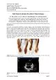
Dr Cróchán OSullivan MD PHD FESC Cardiology service Noninvasive cardiology Advanced Cardiovascular Imaging Transthoracic echo Transoesophageal echo Coronary CT calcium score CT coronary angiography ID: 909734

cardiology coronary chest patients coronary cardiology patients chest pain acute heart pathways invasive implantation rise ecg initial therapy medical

Share:

Link:

Embed:

Download Presentation from below link

Download Presentation The PPT/PDF document "Cardiology Pathway Bon Secours Hospital,..." is the property of its rightful owner. Permission is granted to download and print the materials on this web site for personal, non-commercial use only, and to display it on your personal computer provided you do not modify the materials and that you retain all copyright notices contained in the materials. By downloading content from our website, you accept the terms of this agreement.


Presentation Transcript

Slide1

Cardiology Pathway

Bon Secours Hospital, Cork.

Dr

Cróchán

O’Sullivan MD, PHD, FESC

Slide2

Cardiology service

Slide3

Non-invasive cardiology

Slide4

Advanced Cardiovascular Imaging

Transthoracic echo

Transoesophageal echoCoronary CT calcium scoreCT coronary angiographyStructural Cardiac CTCardiac MRIPerfusion MRIStructural MRI

Slide5

Interventional cardiology

Percutanous

coronary interventionFractional flow reserve (FFR)Instantaneous free-wave ratio (iFR)Intravascular lithotripsyRotaPro

Intracoronary imagingOptical coherence tomographyIntravascular ultrasoundPFO/ASD closureLeft atrial appendage occlusionTranscatheter aortic valve implantation

Slide6

Electrophysiology

Electrophysiological studies.

Ablation of accessory pathways.Pulmonary vein isolation. Atrial flutter ablation. Loop recorder implantation.Permanent pacemaker implantation.Intracardiac defibrillator implantation.Biventricular pacemaker implantation.

Slide7

Natural History of chronic coronary syndromes

Slide8

Initial diagnostic approach for patients with suspected angina

Slide9

Pretest probability of Obstructive Coronary artery disease

Based on a pooled analysis of contemporary data of 15,815 symptomatic patients according to age, sex and the nature of symptoms.

Slide10

Determinants of the clinical likelihood of obstructive coronary artery disease

Slide11

Main diagnostic pathways in symptomatic patients with suspected obstructive coronary artery disease

Slide12

Ischemia trial

nejm

2020

N

Engl

J Med 2020;382:1395-407

5,179 patients with

moderate to severe ischemia

randomized to an

initial invasive strategy

versus an

initial conservative strategy

of medical therapy alone and angiography if medical therapy failed.

Primary endpoint:

composite of

cv death, MI, hospitalization for unstable angina, heart failure or resuscitated cardiac arrest

.

Slide13

Lifestyle management of chronic coronary syndromes

Slide14

Medical therapy of chronic coronary syndromes

Slide15

Decision Tree for patients undergoing invasive coronary angiography

Slide16

Acute

Coronary

Syndrome

1,2

0 h/1 h

hs-Troponin

I-

Algorithm

Acute

Chest

Pain

ECG

hs-Troponin

I

On

admission

and

after 1

hour

.

STEMI-Protocol

Invasive

management

<5

ng

/L

and

rise

in

1

hr.

<2

ng

/L

<2

ng

/L

2-5

ng

/L

and

rise

in

1

hr.

2-5

ng

/L

≥52

ng

/L

5-51

ng

/L

and

rise

in

1

hr.

<6

ng

/L

Rise

in

1

hr.

≥6

ng

/L

Observe

Heart

attack

ruled

out

Heart

attack

probable

Another

test

3

hours

after

admission

Rise

≥10

ng

/L

Pain

free

, Differential

diagnoses

ruled

out

Discharge

/Stresstest

Invasive

investigation

1.

Roffi

M et al. ESC Non-STEMI Guidelines

Eur

Heart J 2015; 2. Reichlin T et al. CMAJ 2015;187(8):E243-E252

ST-Elevation

Ischaemic

ECG

changes

and

persistent

chest

pain

No

Yes

Slide17

Cardiology Referral Pathways

30/06/20

Slide18

Acute Cardiology in the MAU

Prof. Ronan O’SullivanConsultant in Emergency Medicine

Bon Secours Hospital, Cork

Slide19

Cardiology Referral Pathways

Slide20

Chest PainChest pain is high-frequency, high-risk chief complaint

ACS (STEMI/NSTEMI/UA) v ‘low-risk chest pain’Which is more challenging?

Slide21

Clinical HistoryNo one element sufficiently sensitive

Typical v less typical1 in 20 diagnosed w/ acute MI will present atypically

Clinician gestalt very sensitive

Slide22

ECGACEP: Door-to-ECG time 10 minsEven among patients who ultimately rule in for MI, an initial ‘diagnostic’ ECG more exception than rule

ACEP: patients w/ suspected ACS, ECG should be repeated at interval of 30 and 60 mins

Slide23

BiomarkersEssential element of acute assessment

Timing of symptoms relative to biomarker measurementHours/days/constant – single test possible

Intermittent – biomarker stopwatch reset?Not all +ve troponins represent ACS (CHF, renal failure, sepsis)

Slide24

Slide25

Additional TestingEST relatively low yield

Is stress testing still ‘standard of care’? Helpful in patients w/ higher pretest probability of CAD

CT Coronary Calcium Screen/ScoreCoronary CT AngiographyCardiac Perfusion MRIEchocardiographyNon-Cardiac CTPA

Abdominal imaging (USS/CT) Spinal imaging (MRI)

Slide26

Other Causes of Chest PainRespiratory VTE

InfectionGI

MSK (incl. spinal)SkinAnxiety/Stress

Slide27

Cardiology Referral Pathways

Slide28

Other Acute CardiologyAcute/subacute Heart Failure (cave COVID)Palpitations

AF/flutterCollapse query causeSuspected TIA

PresyncopeEcho, Holter/Telemetry, MRI Brain, Carotid USS, CTPA/CXR, bloods, Cardiology review, +/- Neurology review

Slide29

Experience to DateIncrease in chest pain referralsAnxiety-related symptoms (chest pain, dysfunctional breathing)CAD, VTE amongst these…

FTCC referrals redirected to MAUCollapse query causeSuspected TIATGA (transient global amnesia)

Myositis with chest pain

Slide30