/
Diabetic Foot Infection Diabetic Foot Infection

Diabetic Foot Infection - PowerPoint Presentation

jane-oiler
jane-oiler . @jane-oiler
Follow
431 views
Uploaded On 2017-12-15

Diabetic Foot Infection - PPT Presentation

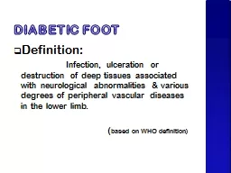
Of all the late complications of diabetes foot problems are probably the most preventable Joslin who wrote in 1934 that diabetic gangrene is not heavensent but earthborn Approximately 5 to 10 of diabetic patients have had past or present foot ulceration and 1 have undergone amputa ID: 615486

infection foot grade diabetic foot infection diabetic grade patients species diabetes wound osteomyelitis ulcer bone neuropathy vascular gangrene gram

Share:

Link:

Embed:

Download Presentation from below link

Download Presentation The PPT/PDF document "Diabetic Foot Infection" is the property of its rightful owner. Permission is granted to download and print the materials on this web site for personal, non-commercial use only, and to display it on your personal computer provided you do not modify the materials and that you retain all copyright notices contained in the materials. By downloading content from our website, you accept the terms of this agreement.


Presentation Transcript

Slide1

Diabetic Foot InfectionSlide2

Of all the late complications of diabetes, foot problems are probably the most preventable.

Joslin

, who wrote in 1934 that “diabetic gangrene is not heaven-sent, but earth-bornSlide3

Approximately 5% to 10% of diabetic patients have had past or present foot ulceration, and 1% have undergone amputationSlide4

Foot infections

Foot infections are among the

most common and serious complications of

diabetes mellitus.

Types of infection include

cellulitis

,

myositis

, abscesses, necrotizing

fasciitis, septic arthritis, tendinitis, and

osteomyelitis

.Slide5

Pathophysiology

Patients with diabetes are particularly susceptible

to foot infection primarily because

of neuropathy, vascular insufficiency, and

diminished

neutrophil

function.

Peripheral

neuropathy has a central role in the development

of a foot infection and it occurs in

about 30 to 50 percent of

patientswith

diabetes.Slide6
Slide7
Slide8

Microbiology

The most common pathogens in acute, previously

untreated, superficial infected foot

wounds in patients with diabetes are aerobic

gram-positive bacteria, particularly

Staphylococcus

aureus

and beta-hemolytic streptococci

(group A, B, and others).Slide9

Microbiology

Infection

in patients who have recently received antibiotics

or who have deep limb-threatening

infection or chronic wounds are

usuallycaused

by a mixture of aerobic gram-positive, aerobic

gram-negative (e.g.,

Escherichia coli, Proteus species,

Klebsiella

species), and anaerobic organisms (e.g.,

Bacteroides

species,

Clostridium species,

Peptococcus

and

Peptostreptococcus

species).Slide10

Key Components of the Comprehensive Diabetic Foot Examination

Dermatologic

   Skin status: color, thickness, dryness, cracking

   Sweating

   Infection: check between toes for fungal infection

   Ulceration

   Calluses/blistering: hemorrhage into callus?Slide11

Musculoskeletal

   Deformity (e.g., claw toes, prominent metatarsal heads, Charcot joint)

   Muscle wasting (guttering between metatarsals)

   Assess whether shoes are appropriate for the feet (e.g., size, width)Slide12

Neurologic

   Ability to perceive pressure from a 10-g monofilament plus one of the following:    Vibration using 128-Hz tuning fork

   Pinprick sensation

   Ankle reflexes

   Vibratory perception thresholdSlide13

VASCULAR

Foot pulses

   Ankle-brachial index, if indicatedSlide14

Clinical Evaluation

existence, severity, and extent of infection, as well as vascular status, neuropathy, and

glycemic

control should be assessed in patients with a diabetic foot infection.

Visible bone and palpable bone on probing are suggestive of underlying

osteomyelitis

in patients with

a diabetic foot infection.

Before an infected wound of a diabetic foot infection is cultured, any overlying necrotic debris should

be removed to eliminate surface contamination and to provide more accurate results.

Routine wound swabs and cultures of material from sinus tracts are unreliable and strongly discouraged

in the management of diabetic foot infection.

The empiric antibiotic regimen for diabetic foot infection should always include an agent active against

Staphylococcus

aureus

, including

methicillin

-resistant S.

aureus

if necessary, and streptococci.Slide15

Meggitt

–Wagner classification of

foot ulcers

Grade Description of the ulcer

Grade 0

Pre- or post-ulcerative lesion

completely

epithelialized

Grade 1

Superficial, full thickness ulcer

limited to the dermis, not

extending to the

subcutis

Grade 2

Ulcer of the skin extending

through the

subcutis

with

exposed tendon or bone and

without

osteomyelitis

or

abscess formation

Grade 3

Deep ulcers with

osteomyelitis

or abscess formation

Grade 4

Localized gangrene of the toes

or the forefoot

Grade 5

Foot with extensive gangreneSlide16

CONFIRMING THE DIAGNOSIS

must be diagnosed clinically rather

than

bacteriologically

The clinical diagnosis of foot infection is

basedon

Thepresence

of purulent discharge from an

ulcer or the classic signs of inflammation (i.e.,

erythema

,

pain, tenderness, warmth, or

induration

). Other suggestive

features of infection include foul odor, the presence

of necrosis, and failure of wound healing despite optimal

managmentSlide17
Slide18
Slide19
Slide20
Slide21
Slide22

Arterial inflow is adequate

   Infection is treated appropriately.

  Pressure is removed from the wound and the immediate surrounding area.Slide23
Slide24
Slide25
Slide26