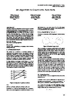
/
International Journal of Computer Applications (0975 International Journal of Computer Applications (0975

International Journal of Computer Applications (0975 - PDF document

jane-oiler
jane-oiler . @jane-oiler
Follow
392 views
Uploaded On 2015-10-29

International Journal of Computer Applications (0975 - PPT Presentation

x2013 8887 Volume 3 x2013 No 7 June 2010 19 Spermatozoa Segmentation and Morphological Parameter Analysis Based Detection of Teratozoospermia VSAbbiramy Dr V Shanthi ID: 176284

– 8887) Volume – No. 7 June

Share:

Link:

Embed:

Download Presentation from below link

Download Pdf The PPT/PDF document "International Journal of Computer Applic..." is the property of its rightful owner. Permission is granted to download and print the materials on this web site for personal, non-commercial use only, and to display it on your personal computer provided you do not modify the materials and that you retain all copyright notices contained in the materials. By downloading content from our website, you accept the terms of this agreement.


Presentation Transcript

International Journal of Computer Applications (0975 – 8887) Volume 3 – No. 7 , June 2010 19 Spermatozoa Segmentation and Morphological Parameter Analysis Based Detection of Teratozoospermia V.S.Abbiramy Dr. V. Shanthi Affiliated to Anna University Affiliated to Anna University Velammal Engine ering College St. Joseph Engineering College Chennai 66 Chennai - 96 ABSTRACT An important parameter as sessed during the semen analysis is the overall morphology, or shape of the sperm . Currently, the morphological analysis of sperm is done manually and is based on visual observation of at least 200 spermatozoa in a microscope followed by a classification s tage based on s trict criteria. But th is method has led to incorrect results due to various factors such as different staining procedures, experience of technicians and human errors. So this paper focuses on morphological classification of spermatozoon eith er as normal or abnormal using Matlab. The first stage is the image preprocessing stage which involves the conversion of RGB image to a gray scale image and then image noises are removed using median filter. The second stage is the detection and extraction of individual spermatozoon which involves the extraction of sperm objects from images using sobel edge detection algorithm. The third stage segments the spermatozoon into various region of interest such as sperm head, midpiece and tail. The fourth stage i nvolves the statistical measurement of spermatozoon which classifies Spermatozoa as normal or abnormal. Keywords : Morphology , Spermatozoon , S permatogenesis , Segmentation, IVF, IUI, WHO , Semen analysis, DNA, Oocyte, Acrosome , Teratozoospermia . 1. INTRODU CTION Infertility is commonly defined as the lack of pregnancy following 12 months of unprotected intercourse. Sperms are produced by a highly complex process of spermatogenesis. Spermatogenesis is the process by which male spermatogonia develop into matur e spermatozoa also known as a sperm cell The basic test of a man's ability to conceive children is the semen analysis. A semen analysis measures certain sperm parameters like sperm count, motility, morphology, volume, fructose level and pH. The current be lief is that sperm morphology assessment should be used primarily as a fertility tool. The measurement of the percentage of spermatozoa having an „ideal‟ morphology using so - called strict method is the method recommended in the latest edition of the World Health Organization (WHO) laboratory manual for semen analysis [ 1]. The shape of the sperm is a reflection of proper sperm development in the testicle, or spermatogenesis. Men with a defect in sperm maturation tend to have problems with sperm morphology a nd may then be at risk for failure of their sperm to fertilize their partner's eggs [ 2] . Strict morphologic assessment was developed to predict fertilization outcomes during In Vitro Fertilization (IVF) and embryo transfer . A positive correlation was foun d between the fertilization rate (FR%) and the proportion of the sperm with a normal (oval) head shape (P .001), the sperm exhibiting acrosomal vacuoles (P .003), the sperm with a normal acrosomal size (40% – 70% ) of total head area (P .025) and the sper m undergoing acrosome reaction after adding human follicular fluid (P .001) [3] . In [4 ] an algorithm for finding sperms in low contrast images was explained. Then, the foreground particles (including sperms and round cells) are segmented from the backgrou nd. Finally, sperms are separated from other cells. The paper [5] h as discussed that neural networks was useful for morphological classification of sperm head . The methodology uses a preprocessing scheme in which invariant Fourier descriptors are lumped into “energy” bands. The resulting networks are pruned using Optimal Brain Damage. T he presence of increased number of morphologically abnormal sperms with impaired motility in males with occupational exposure to high temperature was reported in [ 6]. The author has discussed the relationship between abnormal sperm morphology and chromosomal content or aberrations in individual spermatozoa [7]. In [11] & [12] a novel method for segmenting objects in microscopic images into its constituent's parts is propos ed where t he method called n th fusion is the framewor k of the segmentation algorithm. 2 . SPERM MORPHOLOGY Sperm s are microscopic creatures which look like tiny tadpoles swimming about at a frantic pace. Each sperm is composed of neck, midpiece and tail. The sperm head contains the genetic material of the father in its nucleus. International Journal of Computer Applications (0975 – 8887) Volume 3 – No. 7 , June 2010 20 The mid - piece of t he sperm contains mitochondria, which provide s the energy for sperm motion . The sperm has a long tail in order to propel the head of the sperm, which carries al l the DNA information, towards the egg. A healthy human sperm is about 40 to 250 µ M long and the anatomy of a sperm is shown in the Fig. 2.1 Fig. 2.1 Anatomy of a Sperm Several different shapes or forms of human sperm have been identified and it fall s in to one of the following categories Normal forms Normal sperm have oval head shapes, an intact "mid" section and an uncoiled, single tail as shown in Fig. 2.2 (a). Abnormal heads Some of the sperm head abnormalities are enlarged round head ( Fig. 2.2 (b)) , small head ( Fig. 2.2 (c)) , pinhead ( Fig. 2.2 (d)), double head ( Fig. 2.2 (e)) and an absence of identifiable head are all observed in semen analysis . Taper ed sperm head and constricted head have been seen also . Overall abnormalities in appearance may be termed "amorphous" changes. Abnormal tails Broken tails o r less than half of the normal length should be categorized abnormal. Coiled sperm tail is also sometimes seen as in ( Fig. 2.2 (f)) . Double, triple and quadruple tails are also sometimes seen ( Fig. 2.2 ( g )) and are considered as abnormal. Fig . 2. 2 F rom Top left to right (a) (b) (c) (d) (e) (f) (g) There are two methods for performing sperm morphological eval uations during semen analysis. They are 1. Crude estimation of the percentage of normal sperm specimen 2. Kruger "strict" sperm morphological evaluation. The World Health Organization says good quality semen should contain 60 percent normal sperm morphology. T he strict and WHO morphology score which predicts the sperm's potential for fertilization is shown in Table 2.1 . [ 14]. But most of the labs use " strict” criteria for judging sperm normality . Table 2.1 Strict and WHO III Morphological analysis reference v alues Parameter Percentage of Normal forms Fertilizing capability Strict morphology � 14% Excellent 4 - 14% Decreased 0 - 3% Impaired Fertility or Infertility WHO III Morphology �30% Excellent 15 - 30 Decreased 15% Impaired Fertility 3 . MATERIAL S AND METHODS 3.1 Image Preprocessing This stage is concerned with analyzing the microscopic image and to examine the format of the image. If the image is in RGB form, it is transformed to a gray scale image. The grayscale image is then filtered using med ian filter to remove the noise. The median filter is a nonlinear digital filtering technique , which is often used to remove noise. Matlab 6.5 provides a specialized implementation of the 2D - median filter: K= medfilt2(J); which uses a 3 X 3 neigborhood to compute the median and pads the border of the input with 0s. Applying a 3 X 3 median filter produces an ouput as shown in the Fig. 3.1 International Journal of Computer Applications (0975 – 8887) Volume 3 – No. 7 , June 2010 21 3.2 Detection and extraction of individual spermatozoon Stage 1 : Edges of sperm objects are extracted using S obel e dge detection algorithm. Edge detection is the most common approach for detecting meaningful discontinuities in intensity values. Such discontinuities are detected by using first and second - order derivatives. The first - order derivative of choice in image proce ssing is the gradient. The gradient at the center point in a neighborhood is computed as = = Stage 2: The image is then smoothened to reduce the number of connected components using conv2. C = conv2(A,B) which computes the two - dimensional convolution of matrices A and B . Stage 3: The number of conne cted components in a binary image is calculated using bwlabel. [L, num] = bwlabel (f, conn) where f is an input binary image and conn specifies the desired connectivity (either 4 or 8). Output L is called a label matrix and num gives the total number of connected components found. Stage 4: Any connected components can be extracted using find () . [row, col] = find(X, L==1 ) and the n the extracted spermatozoon is stored in an array which is then displayed . Fig. 3.1 also shows the sample image with abnormal spermatozoa and an extracted abnormal spermatozoon . Fig. 3.2 shows the sample image with normal spermatozoa and an extracted normal spermatozoon. Fig . 3.1 Image with abnormal spermatozoa and an extracted abnormal spermatozoon. Fig. 3. 2 I mage with normal spermatozoa and an extracted normal spermatozoon 3.3 Image Segmentation The main purpose of the segmentation stage is to subdivide a spermatozoon into various constituent parts such as head, mid - piece and tail. Segmentation is done using the Marke r - Controlled W atershed Segmentation. A marker is a connected component belonging to an image. Internal markers and external markers are then computed transform and it follows the following basic procedure: Step 1: Read the color image and convert it to gra yscale. Step 2: Use the gradient magnitude as the segmentation function. Step 3: Compute internal markers which are inside each of the objects of interest. Step 4: Compute external markers which are contained within the background. These are pixels that are not part of any object. Step 5: Use internal and external markers to modify the gradient image by a procedure called minima imposition. It modifies a gray - scale image so that regional minima occur only in marked locations. Other pixels are pushed up as necessary to remove all other regional minima. Step 6: Finally, compute the watershed transform of the marker - modified gradient image a nd t he result s are shown in Fig. 3.3.1, Fig 3.3.2 & Fig. 3.3.3 Fig. 3.3.1 Result of the Image Segmentation stage International Journal of Computer Applications (0975 – 8887) Volume 3 – No. 7 , June 2010 22 Fig. 3.3.2 Result of the Image Segmentation stage Fig. 3.3.3 Result of the Image Segmentation stage 3.4 FEATURE EXTRACTION Feature extraction is performed over the image extracted from the previous stage. These features are used to classify H ead Area: It is the number of pixels contained in the segmented head region. Perimeter: It is the number of pixels in the boundary of the Spermatozoa. Head Length: Head length (in pixels) is calculated with the major axis. Major axis is defined as a l ine that contains the center of mass point and has a slope equal to the line defined by the highest value pixel in an Euclidean distance transform. Head width: Head width (in pixels ) is calculated with the minor axis. Minor axis is defined as the line perp endicular to the major axis . Mid - piece length: It is the major axis of the midpiece of the mid - piece and it is measured in pixels. Tail Length: It is the major axis of the midpiece of the tail and it is measured in pixels. Orientation : It is the angle (in degrees) between the neck and tail to the major axis of the head. Eccentricity: The ratio of the head length to the head width (ie.major axis to the minor axis ) is called the eccentricity. Equvidiameter : It is defined as the Euclidean distance between t he two farthest points on the boundary. 4. RESULTS AND ANALYSIS The Table 4.1 shows the statistical measurements of morphological parameters of spermatozoon obtained based on which the images are classified as Normal or Abnormal spermatozoon. 5. CONCLUSIO NS AND FUTURE WORK Even though the assessment of sperm morphology for either normal spermatozoa or for sperm defects is relatively extensive, the different stages in the analysis of human spermatozoon morphology were presented. This paper has focused on th e measurement of parameters like sperm head length, width, area, perimeter, midpiece length and tail length. It is possible to detect the sperm abnormalities such as round heads, pin heads, very large heads, double heads, abnormal midpiece, absent tails a nd double tails. As abnormal sperm will not be able to fertilize the egg, morphologic assessment discussed in this paper could be helpful to detect the pregnancy outcomes in couples undergoing Intrauterine Insemination / In Vitro Fertilization ( IUI/IVF). As the size and shape of the acrosome is particularly important for sperm binding to the oocyte, this paper could be extended to measure the level of acrosome in the sperm head. „Excess residual cytoplasm‟ on sperm produced by imperfect sperm atogenesis cou ld also to be measured. The mitochondria in the midpiece supplies the energy for the sperm's activity, it needs to be analysed. ACKNOWLEDGMENTS I extend my sincere thanks to the reviewers for their valuable comments. International Journal of Computer Applications (0975 – 8887) Volume 3 – No. 7 , June 2010 23 Table 4.1 Statistical Measurement of Morphological Parameters of Spermatozoon Courtesy: The images used in this paper algorithm were obtained from the Advanced Fertility Center of Chicago, Infertility and In Vitro Fertilization with ICSI Specialists , Gurnee & Crystal Lake, Illinois. Fixed and stained human sperm pictures with high magnification are taken from IVF lab. 6. REFERENCES [1] Assessing human sperm morphology: top models, underdogs or biometrics? Jacques Auger, Asian Journal of Andrology (2010) 12: 36 – 46. [2] http://babymed.com/FAQ/Content.aspx?14619 [3] The Human Sperm Head: A Key for Successful Fertilization, Alaa A. El - Ghobashy And Christopher R. West, Journal of Andrology, Vol. 24, No. 2, March/April 2003 [4] Sperm Ident ification Using Elliptic Model and Tail Detection, Vahid Reza Nafisi, Mohammad Hasan Moradi, and Mohammad Hosain Nasr - Esfahani, World Academy of Science, Engineering and Technology 6 2005 [5] Towards Semen Quality Assessment Using Neural Networks, Chr. Li nneberg, P. Salamon, 0 - 7803 - 2026 - 3/94 , 1994 IEEE [6] Deterioration Of Sperm Morphology In Men Exposed To High Temperature, Dada R., Gupta, N.P., Kucheria K., J Anat. Soc. India 50(2) 107 - 111 (2001) [7] Human Sperm Maintain Their Shape Following Deconden sation and Denaturation for Fluorescent In Situ Hybridization: Shape Analysis and Objective Morphometry1, Ciler Celik - Ozenci , Biology Of Reproduction 69, 1347 – 1355 (2003) [8] http://www.gfmer.ch/Endo/PGC_network/Sperm_ morphology.htm [9] The human acroso me reaction, H.W.G. Baker, D.Y. Liu, C. Garrett, M. Martic , University of Melbourne Department of Obstetrics and Gynaecology, Asian J Androl 2000 Sep; 2: 172 - 178 [10 ] WHO laboratory manual for examination of human semen and sperm cervical mucus interact ion. 4 Ed. Cambridge: Cambridge university press, 1999. [11] Spermatozoon Segmentation Towards an Objective Analysis of Human Sperm Morphology, , Henry Carrillo et al., Department of Electrical and Electronic Engineering, Colombia, Proceedings of the 5t h International Symposium on image and Signal Processing and Analysis (2007) [12] A Computer Aided Tool for the Assessment of Human Sperm Morphology, Henry Carrillo et al., Department of Electrical and Electronic Engineering, Colombia, 1 - 4244 - 1509 - 8/07/$ 25.00 02007 IEEE [13] Digital Image Processing Using Matlab, Gonzales, Rafael. Et al, New Jersey, Pearson Education, 2004. [14] http://www.umc.sunysb.edu/urology/male_infertility/ SEMEN_ANALYSIS.html Input Images Area Perimeter Sperm Head Eccentricity Midpiece Length Tail Length Orientation Equiv Diameter Class Length Width Image1 36 46.7032 9.8914 5.3217 0.8429 15.62 67.57 78.5457 6.770 3 Normal Image2 158 191.2335 23.6182 8.6646 0.9303 19.11 66.23 81.9619 13.9116 Abnormal (Large Head) Image3 1 3.6280 1.1547 1.1547 0.9600 10.23 44.69 0 1.1284 Abnormal (Smaller Head) Image4 37 25.453 10.1335 5.3444 0.8496 19.12 35.54 88.9338 6.86 37 Abnormal ( bent midpiece) Image5 10 11.8454 4.2583 3.2083 0.6575 14 74.31 90 3.5682 Abnormal Image6 1 3.6280 1.1547 1.1547 0 15.07 44.16 0 1.1284 Abnormal (Pinheaded) Image7 7 13.4457 5.7735 1.8145 0.9493 15.35 38.77 45 2.9854 Abnormal ( Do uble Tail) Image8 7 10.1697 3.8791 2.4300 0.7795 15.35 42.27 45 2.9854 Abnormal ( Double Headed)