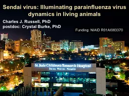
/
Sendai virus: Illuminating parainfluenza virus dynamics in Sendai virus: Illuminating parainfluenza virus dynamics in

Sendai virus: Illuminating parainfluenza virus dynamics in - PowerPoint Presentation

jane-oiler
jane-oiler . @jane-oiler
Follow
440 views
Uploaded On 2016-12-09

Sendai virus: Illuminating parainfluenza virus dynamics in - PPT Presentation

Charles J Russell PhD postdoc Crystal Burke PhD Funding NIAID R01AI083370 HPIV1 HPIV2 HPIV3 leading cause of pediatric hospitalization 21000 yr in USA virtually all infected by age 5 reinfections common but usually less severe ID: 499604

transmission infection russell virus infection transmission virus russell pfu contact burke

Share:

Link:

Embed:

Download Presentation from below link

Download Presentation The PPT/PDF document "Sendai virus: Illuminating parainfluenza..." is the property of its rightful owner. Permission is granted to download and print the materials on this web site for personal, non-commercial use only, and to display it on your personal computer provided you do not modify the materials and that you retain all copyright notices contained in the materials. By downloading content from our website, you accept the terms of this agreement.


Presentation Transcript

Slide1

Sendai virus: Illuminating parainfluenza virus dynamics in living animals

Charles J. Russell, PhD

postdoc: Crystal Burke, PhD

Funding: NIAID R01AI083370Slide2

HPIV1, HPIV2, HPIV3 leading cause of pediatric hospitalization (21,000/yr in USA)

virtually all infected by age 5; reinfections common but usually less severe

no available anti-PIV drugs or vaccinesParamyxoviruses replicate in epithelial cells that line the respiratory tract, causing inflammation in the nasopharynx

,

larnyx, trachea & lungsImportant causes of croup (laryngotracheobronchitis) and pneumonia

Human parainfluenza virusesSlide3

Cross-protective immune responses (Jennerian vaccine)

Tracheal infection/inflammation (croup)

Efficient contact transmission

Reinfection can occur

Majority of healthy hosts do not suffer severe LRT infection

Lamb &

Kolakofsky

, 2001

Fields Virology

Sendai virus: murine counterpart of HPIV1Slide4

N

P

M

F

HN

L

luciferase

WT-like reporter virus: MF*

optimize gene start sequence

Burke…Russell 2011 PLoS PathogensSlide5

Imaging infection daily in a living mouse

1

2

9

8

7

6

5

4

3

10

7000

PFU

M-F

*

in 30

μl

day:

lungs

highest

lowest

Burke…Russell 2011 PLoS PathogensSlide6

Bioluminescence in

Nasopharynx

Bioluminescence in Lungs

Weight Change

Resistant in lungs but susceptible in URT

Burke…Russell 2011 PLoS PathogensSlide7

Nasopharynx

Lungs

Weight loss

Low-dose inoculation grows to high level in URT

7000 PFU

70 PFU

Burke…Russell 2011 PLoS PathogensSlide8

day

post

infection

70 or 7000 PFU,

BALB/

c or 129 mice

0

1

14

luminescence

1º infection or

transmission

76

71

70

luminescence

reinfection

3x10

6

PFU

challenge

30

Contact transmissionSlide9

Contact transmission70 PFU or7000 PFU virus

“resistant” BALB/c

“susceptible” 129 mice100% contact transmission

similar-looking URT-biased infection in recipients

protects from lethal challengeSlide10

1.

Nasopharynx

2. Trachea (~0.8 days later)

3. Lungs (~1.0 days later)

For both 129/SvJ and BALB/c mouse strains

and 70- or 7,000-PFU inoculations into donors

highest

lowest

Progression of 1° infection in contact recipient miceSlide11

3.4 days

3.3 days

7,000 PFU inoculation

Susceptibility to lung infection does not affect contact transmission.

Nasal virus shedding in inoculated mice => contact transmission.

Time until detection in

nasopharynxSlide12

Looks like a low-dose, low-volume, URT-biased infection

nasopharynx

trachea

lungs

70

PFU in 5

m

L

Contact transmission

Contact transmission

Burke…Russell 2013 PLoS PathogensSlide13

donors

isolated

recipients

Air flow

129-strain

“susceptible”

mice

7.6

or

15

cm

Airborne transmission

day

of

expt.

0

1

14

primary

76

71

70

challenge

30

3x10

6

PFU

Burke…Russell 2013 PLoS PathogensSlide14

(4/21)(5/21)

(8/21)

Working hypothesis: Dynamics of infection determined by

the

site of inoculation & infectious doseDiverse dynamics of primary infection after airborne transmission

day: 1 2 3 4 5 6 7 8 9 10 11 12 13 14 15

Burke…Russell 2013 PLoS PathogensSlide15

1° infection inversely correlates with reinfection

Nasal first

4/21

No

transmission

3/21

Tracheal dominant

8/21

Tracheal first

5/21

Burke…Russell 2013 PLoS PathogensSlide16

Protection from natural reinfection by contact transmission

Intranasal vaccination with a low dose/volume of attenuated virus: no reinfection.

Intramuscular vaccination: reinfection in the nasopharynx and trachea.

Burke…Russell 2014 submittedSlide17

Decoupling of Sendai virus infection in upper versus lower respiratory tract Lung infection and concomitant host response determines pathogenesis

Upper respiratory tract infection determines transmission & induces protective immunity even under suboptimal conditions

Clinical diagnosis: titers from nasal washes not same as lung titers

Vaccine development

: attenuated or lower-dose I.N. live-virus vaccinesParadigm for respiratory virus infection: for a virus matched to its host, ‘natural’ infection after transmission elicits immunity without pathology Robust upper respiratory tract infection benefits both virus and the host

Mode of transmission determines the tropism and magnitude of primary infection, which is in turn inversely correlated with reinfection

ANISOTROPIC INFECTIONS: Dynamics of natural respiratory infections can vary. Compartmentalization of immune response contributes to protection from reinfectionMajor FindingsSlide18

Sendai virus: Illuminating parainfluenza virus dynamics in living animals

Charles J. Russell, PhD

postdoc: Crystal Burke, PhD

Funding: NIAID R01AI083370