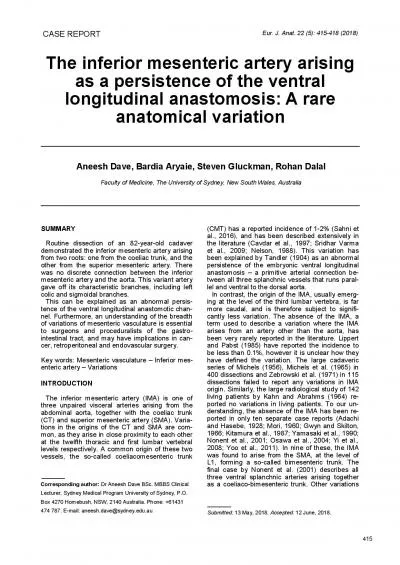
/
Capillaries Capillaries _Are very small vessels, their diameter ranges from (4-15)Mm, Capillaries Capillaries _Are very small vessels, their diameter ranges from (4-15)Mm,

Capillaries Capillaries _Are very small vessels, their diameter ranges from (4-15)Mm, - PowerPoint Presentation

joanne
joanne . @joanne
Follow
64 views
Uploaded On 2024-01-29

Capillaries Capillaries _Are very small vessels, their diameter ranges from (4-15)Mm, - PPT Presentation

The wall of a segment of capillarity may be formed by a single endothelial cell Only tunica intimae is present which typically consist of the endothelium its basal lamina and an in complete layer of cells surrounding the capillary the pericytes ID: 1041427

cells capillaries vessels endothelial capillaries cells endothelial vessels pericytes blood continuous cap wall phagocytic muscle capillary fenestrated present type

Share:

Link:

Embed:

Download Presentation from below link

Download Presentation The PPT/PDF document "Capillaries Capillaries _Are very small ..." is the property of its rightful owner. Permission is granted to download and print the materials on this web site for personal, non-commercial use only, and to display it on your personal computer provided you do not modify the materials and that you retain all copyright notices contained in the materials. By downloading content from our website, you accept the terms of this agreement.


Presentation Transcript

1. Capillaries

2. Capillaries_Are very small vessels, their diameter ranges from (4-15)Mm, arising from the terminal end of the arterioles. _The wall of a segment of capillarity may be formed by a single endothelial cell. _Only tunica intimae is present ,which typically consist of the endothelium, its basal lamina and an in complete layer of cells surrounding the capillary, the pericytes.

3. These pericytes appear to have important roles in repair of blood vessels and connective tissue after injury, these cells have contractile properties and can regulate blood flow in capillaries. They have the potential to develop in to fibroblasts, smooth muscle cells and may even be phagocytic.

4. 1-Continouous capillaries (type I): The endothelial cells forms a continuous internal lining without any intercellular or intra cytoplasmic defects, pericytes may be found in a associated with the endothelium of certain continuous capillaries. Found in muscle,lung,CNS.Classification of Capillaries

5.

6. 2-Fenestrated Capillaries (type II):The endothelial cells are pierced by pores, which provide channels across the capillary wall.Found in GIT mucosa, endocrine glands and in the renal (glomeruli).3-Discontinuous capillaries (Sinusoid):Are larger and more irregularly shaped than other capillaries, they are thin walled blood vessels lined by endothelial cells and specialized phagocytic cells, found in liver,spleen,bon marrow. usually wide gabs are present between the endothelial cells that permit leakage of material in to and out of these vessels.

7.

8.

9. continuous cap.fenestrated cap.sinusoidClassifications and ultrastrutural feature of Cap.