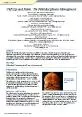
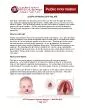
OROFACIAL CLEFTS 1CLEFT LIP AND PALATE Cleft lip It is a developmental anomaly characterized by a wedgeshaped defect in the lip which results from failure of two parts of the lip to fuse together at the time of development ID: 912540
Download Presentation The PPT/PDF document "Developmental Defects of the Lip and P..." is the property of its rightful owner. Permission is granted to download and print the materials on this web site for personal, non-commercial use only, and to display it on your personal computer provided you do not modify the materials and that you retain all copyright notices contained in the materials. By downloading content from our website, you accept the terms of this agreement.
Slide1
Developmental Defects of the Lip and Palate
Slide2OROFACIAL CLEFTS
• 1-CLEFT LIP AND PALATECleft lip: It is a developmental anomaly characterized by a wedge-shaped defect in the lip, which results from failure of two parts of the lip to fuse together at the time of development. This defect is more commonly seen in relation to the upper lip.
Slide3CLEFT PALATE
It is a developmental defect of palate characterized by lack of complete fusion of two lateral halves of the palate resulting in a cleft. Cleft in the palate leads to communication between oral and the nasal cavity.
Slide4ETIOLOGY:
Heredity. Environmental factors such as: Insufficent nutrition to pregnant women Defective vascular supply Size of the tongue prevent union of affected parts Infections , certain alcohol ,drugs and toxins
Slide5Classification
• cleft lip • Unilateral (usually on the left side), with or without an anterior alveolar ridge cleft • Bilateral, with or without alveolar ridge clefts, complete or incomplete Palatal clefts • Bifid uvula
•
Soft palate only
•
Both hard and soft palate
Slide6Combined lip and palatal defects
• Unilateral, complete or incomplete • Cleft palate with bilateral cleft lip, complete or incomplete
Slide7Oblique Facial Cleft
Extends from the upper lip to the eye. It is nearly always associated with CP.Some of these clefts may represent failure of fusion of the lateral nasal process with the maxillary process.
Slide8Lateral Facial cleft
It is caused by lack of fusion of the maxillary and mandibular processes. This cleft may be unilateral or bilateral.Extending from the commissure toward the ear, resulting in macrostomia. May occur as an isolated defect, but more often it is associated with other disorders.
Slide9Slide10Slide11Slide12Congenital lip pits
Slide13Slide14Commissural lip pits
Slide15Developmental Defects of the Jaw Bones
Slide16AGNATHIA
Absence of one of the jawsRare conditionMostly occurs in mandible
Slide17MACROGNATHIA
Abnormally large jawDue to :- Fibrous dysplasia- Bone tumors - Odontogenic cysts- Associated with acromegaly or pagets disease
Slide18MICROGNATHIA
Abnormally small size of one of the jawsMay be associated with Pierre Robin syndrome
Slide19coronoid
HyperplasiaA rare developmental anomaly that may result in limitation of mandibular movement. The cause of coronoid hyperplasia is unknown. Because most cases have been seen in pubertal males, an endocrine influence has been suggested. Coronoid hyperplasia may be unilateral or bilateral,
Unilateral enlargement of the
coronoid
process also can result from a
true tumor, such as an
osteoma
or
osteochondroma
,.
Slide20Condylar
HyperplasiaExcessive growth of one condyleUnknown causeEndocrine disturbances and trauma may be etiological factors
Slide21Hemifacial
HypertrophyUnilateral enlargement of face as a result of increased neurovascular supply of to the affected side of the face resulting in A symmetry of the face , malocclusion, deviation f the affected side of face to unaffected one.
Slide22Hemifacial
AtrofyUnknown etiologyAtrophic changes affecting one side of the face.Mouth and nose are deviated toward the defective side
Slide23STAFNE DEFECT (STAFNE BONE CYST;LINGUAL MANDIBULAR SALIVARYGLAND DEPRESSION)
Developmental concavity of the cortex of the mandible in the molar area.Formed around an accessory lateral lobe of submandibular glandRadiographic appearance is of well circumscribed cystic lesion within the bone usually below the inferior alveolar canal.Histologically normal salivary gland tissue suggesting that it is developmental defects
Slide24Mandibular
dysostosis (treacher –collins syndrome)Autosomal dominant disorderHypoplastic zygoma resulting narrow face with depressed cheek..
Underdeveloped mandible with
retruded
chin and cleft palate may be seen.
Mandibulofacial
dysostosis
. Patient exhibits a
hypoplastic
mandible.
Slide25Cleidocranial
dysplasia or dysostosisRare familial disorder characterised by defective formation of clavicles sometimes retrusion of maxilla.Delayed eruption of permanent dentition Supernumerary teeth may be seen radigraphically
.
Slide26Condylar
hypoplasia, or underdevelopment of the mandibular condyle Can be either congenital or acquired. Congenital condylar hypoplasia often is associated with head and neck syndromes, including
mandibulofacial
dysostosis
and
hemifacial microsomia
.
Acquired
condylar
hypoplasia
results from disturbances of the growth center of the developing
condyle
.
The most frequent cause is trauma to the
condylar
region during infancy or childhood.
Other causes include infections, radiation therapy, and rheumatoid
arthritis.
Slide27Slide28Bifid
condyle It is a rare developmental anomaly characterized by A double-headed mandibular condyle.Most bifid condyles have a medial and lateral head divided by an anteroposterior
groove.
Some
condyles
may be divided into an anterior and posterior head.
The cause of bifid
condyle
is uncertain.
Anteroposterior
bifid
condyles
may be of traumatic origin, such
as a childhood fracture.
Mediolaterally
divided
condyles
may result from trauma, abnormal muscle attachment
Slide29Bifid
condyle. Radiograph of the mandibularcondyle showing a double head (arrow).
Slide30Torus
palatinus Presents as a bony hard mass that arises along the midline suture of the hard palate
Slide31Torus Palatinus
Slide32Tori sometimes are classified according to their morphologic appearance: -The flat torus has a broad base and a slightly convex, smooth surface. It extends symmetrically onto both sides of the midline raphe.-The spindle torus has a midline ridge along the palatal raphe. -The nodular torus arises as multiple protuberances, each with an individual base.
-The lobular torus is also a
lobulated
mass, but it rises from a single base.
Slide33Torus
Mandibularis It is an exostosis covered with normal mucosa that appears on the lingual surfaces of the mandible, usually in the area adjacent to the bicuspids .The incidence of torus mandibularis is about 6%. Bilateral exostoses occur in 80% of the cases. Clinically, it is an asymptomatic growth that varies in size and shape.
Slide34Torus mandibularis
Slide35Bony
Exostoses Multiple exostoses are rare and may occur on the buccal surface of the maxilla and the mandible. Clinically, they appear as multiple asymptomatic small nodular, bony elevations below the muccolabial fold covered with normal mucosa. The cause is unknown and the lesions are benign, requiring no therapy.
Problems may be encountered during denture preparation.