/
World Journal of Pharmaceutical and Medical Research World Journal of Pharmaceutical and Medical Research

World Journal of Pharmaceutical and Medical Research - PDF document

josephine
josephine . @josephine
Follow
345 views
Uploaded On 2021-02-11

World Journal of Pharmaceutical and Medical Research - PPT Presentation

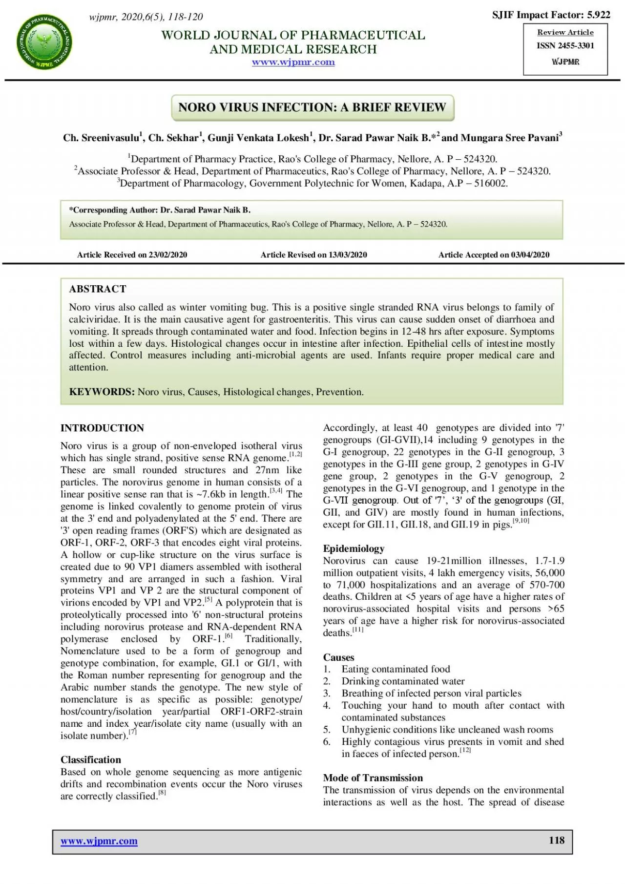
Naik et al wwwwjpmrcom 118 NORO VIRUS INFECTION A BRIEF REVIEW Ch Sreenivasulu 1 Ch Sekhar 1 Gunji Venkata Lokesh 1 Dr Sarad Pawar Naik B 2 a ID: 830905

norovirus virus genotypes journal virus norovirus journal genotypes noro genogroup viral genome medical viruses rna infected disease food contaminated

Share:

Link:

Embed:

Download Presentation from below link

Download Pdf The PPT/PDF document "World Journal of Pharmaceutical and Medi..." is the property of its rightful owner. Permission is granted to download and print the materials on this web site for personal, non-commercial use only, and to display it on your personal computer provided you do not modify the materials and that you retain all copyright notices contained in the materials. By downloading content from our website, you accept the terms of this agreement.


Presentation Transcript

Naik et al.
Naik et al. World Journal of Pharmaceutical and Medical Research www.wjpmr.com 118 NORO VIRUS INFECTION: A BRIEF REVIEW Ch. Sreenivasulu1, Ch. Sekhar1, Gunji Venkata Lokesh1, Dr. Sarad Pawar Naik B.*2 and Mungara Sree Pavani3 1Department of Pharmacy Practice, Rao's College of Pharmacy, Nellore, A. P – 524320. 2Associate Professor & Head, Department of Pharmaceutics, Rao's College of Pharmacy, Nellore, A. P – 524320. 3Department of Pharmacology, Government Polytechnic for Women, Kadapa, A.P – 516002. Article Received on 23/02/2020 Article Revised on 13/03/2020 Article Accepted on 03/04/2020 INTRODUCTION Noro virus is a group of non-enveloped isotheral virus which has single strand, positive sense RNA genome.[1,2] These are small rounded structures and 27nm like particles. The norovirus genome in human consists of a linear positive sense ran that is ~7.6kb in length.[3,4] The genome is linked covalently to genome protein of virus at the 3' end and polyadenylated at the 5' end. There are '3' open reading frames (ORF'S) which are designated as ORF-1, ORF-2, ORF-3 that encodes eight viral proteins. A hollow or cup-like structure on the virus surface is created due to 90 VP1 diamers assembled with isotheral symmetry and are arranged in such a fashion. Viral proteins VP1 and VP 2 are the structural component of virions encoded by VP1 and VP2.[5] A polyprotein that is proteolytically processed into '6' non-structural proteins including norovirus protease and RNA-dependent RNA polymerase enclosed by ORF-1.[6] Traditionally, Nomenclature used to be a form of genogroup and genotype combination, for example, GI.1 or GI/1, with the Roman number representing for genogroup and the Arabic number stands the genotype. The new style of nomenclature is as specific as possible: genotype/ host/country/isolation year/partial ORF1-ORF2-strain name and index year/isolate city name (usually with an isolate number).[7] Classification Based on whole genome sequencing as more antigenic drifts and recombination events occur the Noro viruses are correctly classified.[8] Accordingly, at least 40 genotypes are divided into '7' genogroups (GI-GVII),14 including 9 genotypes in the G-I genogroup, 22 genotypes in the G-II genogroup, 3 genotypes in the G-III gene group, 2 genotypes in G-IV gene group, 2 genotypes in the G-V genogroup, 2 genotypes in the G-VI genogroup, and 1 genotype in the G-VII genogroup. Out of '7’, ‘3' of the genogroups (GI, GII, and GIV) are mostly found in human infections, except for GII.11, GII.18, and GII.19 in pigs.[9,10] Epidemiology Norovirus can cause 19-21million illnesses, 1.7-1.9 million outpatient visits, 4 lakh emergency visits, 56,000 to 71,000 hospitalizations and an average of 570-700 deaths. Children at years of age have a higher rates of norovirus-associated hospital visits and persons �65 years of age have a higher risk for norovirus-associated deaths.[11] Causes 1. Eating contaminated food 2. Drinking contaminated water 3. Breathing of infected person viral particles 4. Touching your hand to mouth after contact with contaminated substances 5. Unhygienic conditions like uncleaned wash rooms 6. Highly contagious virus presents in vomit and shed in faeces of infected person.[12] Mode of Transmission The transmission of virus depends on the environmental interactions as well as the host. The spread of disease wjpmr, 2020,6(5), 118-120 SJIF Impact Factor: 5.922 Review Article ISSN 2455-3301 WJPMR WORLD JOURNAL OF PHARMACEUTICAL AND MEDICAL RESEARCH www.wjpmr.com *Correspo

nding Author: Dr. Sarad Pawar Naik B.
nding Author: Dr. Sarad Pawar Naik B. Associate Professor & Head, Department of Pharmaceutics, Rao's College of Pharmacy, Nellore, A. P – 524320. ABSTRACT Noro virus also called as winter vomiting bug. This is a positive single stranded RNA virus belongs to family of calciviridae. It is the main causative agent for gastroenteritis. This virus can cause sudden onset of diarrhoea and vomiting. It spreads through contaminated water and food. Infection begins in 12-48 hrs after exposure. Symptoms lost within a few days. Histological changes occur in intestine after infection. Epithelial cells of intestine mostly affected. Control measures including anti-microbial agents are used. Infants require proper medical care and attention. KEYWORDS: Noro virus, Causes, Histological changes, Prevention. Naik et al. World Journal of Pharmaceutical and Medical Research www.wjpmr.com 119 mostly occurs in places like schools, hotels, hospitals and nursing homes very quickly. Probably, Viruses are the most common cause of infectious disease acquired indoor.[13] Symptoms  Low grade fever  Nausea and severe vomiting  Loose or watery diarrhoea  Malaise  Body aches  Muscle cramps  Sudden sickness Histological changes Specific histological changes occur in mucosa of intestine such as broadening and blunting of villi and shortening of microvilli, when norovirus interacts with intestinal mucosa. Intracellular oedema, increased cytoplasmic vacuolation and pale and enlarged mitochondria also occurs. Abnormalities can be appeared in epithelial cells present in intestine of norovirus- infected region. Norovirus-infected volunteer electron microscopy analysis shows that these cells remain intact, hyperplasia of crypt cell has also been reported.[14] Mild inflammatory infiltration into the lamina propria intestinal lesions are the enterocyte changes can be seen, which resolves within '2' weeks. Enterocytes apoptosis are also seen in humans, mice and pigs. Enterocyte apoptosis caused by Lymphocytes during Norovirus infection and upon this can cause both direct and indirect mechanism to norovirus induced apoptosis of enterocyte due to release of perforin. This can infect the host for weeks or month. The norovirus present in a fully functional immune system, that becomes the source of virus continuing its spread within the population that is similar to Feline caliciviruses.[15] Diagnosis ELISA and RT-PCR methods are commonly used during outbreaks. The sensitivity of ELISA kits and the Rainscreen test were found to be disappointed. The screening of ELISA is used un preliminary testing. Faecal sample testing is most commonly used for the identification of virus.[16] Prevention  Maintenance of clean environment.  Isolation from infected person.  Clean hand washing frequently.  Well cleaned vegetables and fruits.  Contaminated food and water should be avoided.  Surrounding area should be neat.  Virus containing area must be disinfected.  Vomit or faeces should be cleaned properly.  A broad range of anti-microbial solutions and disinfectants are used for better results.[17] CONCLUSION Noro virus is most detected pathogen in sporadic diseases. Noro virus are pathogenic agents which leads to which leads to acute and chronic gastroenteritis in humans of all ages. There is no specific treatment and recovery depends upon health of the immune system. There is no vaccine against this virus. At present, there are no anti-viral medications to treat the norovirus. Most of the cases recovered without treatment. Alternative care is given by fluids. Drink more amount of water to replace the water lost in dehydration, caused by vomiti

ng and diarrhoea. Special drinks for r
ng and diarrhoea. Special drinks for rehydration can also be administered. The cost of this single nosocomial outbreak support aggressive effort to prevent virus transmission. Preventive measures should be followed to reduce the spread of infection. Personal hygiene and isolation from the infected persons. REFERENCES 1. Koopmans M, Duizer E. Foodborne viruses: an emerging problem. International journal of food microbiology, 2004; 90(1): 23-41. 2. Cohn A, Jackson ML, Brunette GW, Kozarsky PE, Magill AJ, Shlim DR. Meningococcal disease. In CDC health information for international travel 2012. The yellow book, 2012; 250-253. 3. Dennehy PH. Viral gastroenteritis in children. The Paediatric infectious disease journal, 2011; 30(1): 63- 4. 4. Yap J, Qadir A, Liu I, Loh J, Tan BH, Lee VJ. Outbreak of acute norovirus gastroenteritis in a military facility in Singapore: a public health perspective. Singapore medical journal, 2012; 1: 53. 5. La Rosa G, Fratini M, Libera SD, Iaconelli M, Muscillo M. Viral infections acquired indoors through airborne, droplet or contact transmission. Annali dell'Istituto superiore di sanita, 2013; 49(2): 124-32. (4): 249. 6. Le Gall O, Christian P, Fauquet CM, King AM, Knowles NJ, Nakashima N, Stanway G, Gorbalenya AE. Picornavirales, a proposed order of positive-sense single-stranded RNA viruses with a pseudo-T= 3 virion architecture. Archives of virology, 2008; 153(4): 715. 7. Moore MD, Goulter RM, Jaykus LA. Human norovirus as a foodborne pathogen: challenges and developments. Annu Rev Food Sci Technol, 2015; 6: 411-433. doi:10.1146/annurevfood-022814-015643. 8. Hernandez JD, Silva LD, Sousa ECJ, et al. Analysis of uncommon norovirus recombinants from Manaus, Amazon region, Brazil: G-II. P22/G-II.5, G-II. P7/G-II.6 and G-II. Pg/G-II.1. Infect Genet Evol, 2016; 39: 365-371. doi: 10.1016/j.meegid.2016.02.007. 9. Wang QH, Costantini V, Saif LJ. Porcine enteric caliciviruses: genetic and antigenic relatedness to human caliciviruses, diagnosis and epidemiology. Vaccine, 2007; 25(30): 5453-5466. doi: 10.1016/j.vaccine.2006.12.032. Naik et al. World Journal of Pharmaceutical and Medical Research www.wjpmr.com 120 10. Shen Q, Zhang W, Yang S, Cui L, Hua X. Complete genome sequence of a new-genotype porcine norovirus isolated from piglets with diarrhoea. J Virol, 2012; 86(12): 7015-7016. doi:10.1128/JVI.00757-12. 11. Rabenau HF, Stürmer M, Buxbaum S, Walczok A, Preiser W, Doer HW. Laboratory diagnosis of norovirus: which method is the best? Intervirology, 2003; 46(4): 232-8. 12. Ohashi N, Yonemura K, Sugiura T, Isozaki T, Togawa A, Fujigaki Y, Yamamoto T, Hishida A. Withdrawal of interferon-α results in prompt resolution of thrombocytopenia and haemolysis but not renal failure in hemolytic uremic syndrome caused by interferon-α. American Journal of Kidney Diseases, 2003; 41(3): e10-1. 13. Graw F, Perelson AS. Modelling viral spread. Annual review of virology, 2016, 3: 555-72. 14. Chikh Ali M, Karasev AV, Furutani N, Taniguchi M, Kano Y, Sato M, Natsuaki T, Maoka T. Occurrence of Potato virus Y strain PVYNTN in foundation seed potatoes in Japan, and screening for symptoms in Japanese potato cultivars. Plant Pathology, 2013; 62(5): 1157-65. 15. Karst SM. Pathogenesis of noroviruses, emerging RNA viruses. Viruses, 2010; 2(3): 748-81. 16. De Bruin E, Duizer E, Vennema H, Koopmans MP. Diagnosis of Norovirus outbreaks by commercial ELISA or RT-PCR. Journal of virological methods, 2006; 137(2): 259-64. 17. Centres for Disease Control and Prevention (CDC. Norovirus activity--United States, 2006-2007. MMWR. Morbidity and mortality weekly report, 2007; 56(33): 842